We present a case of a female who sustained presumed frostbite over the right cheekbone a few weeks after undergoing dermal filler cheekbone augmentation injections.
To our knowledge, this is the first reported case of focal frostbite over dermal filler thought to be linked to loss of perfusion capability possibly induced by foreign substance filler cheekbone augmentation injections.
The most common anatomic sites of frostbite are the ears, nose, fingers and toes.1,3 8,9 The dermal filler injection elevates the tissue off the bone and occupies space between the tissue and bone.2 As dermal filler facial augmentation injection becomes more mainstream and this population participates in outdoor cold weather activities, an increased risk of cold injury in thin tissue over dermal filler remains possible. Patients undergoing dermal filler augmentation injections should be vigilant about cold injury when adventuring in extreme cold environments.
Case
A 48-year-old female sustained a 2.5 cm x 1.5 cm focal area of presumed frostbite over the right cheekbone. The patient had a history of Restylene Lyft™ cheekbone augmentation performed at the area without incident or complication 58 days prior to cold exposure. The patient is a never-smoker who has no other relevant medical or surgical history and takes no medications beyond over the counter supplements.
The patient had planned a 4-hour backcountry excursion in Wyoming, including snowmobiling, snowshoeing, and hiking. The temperature was -10°F at the start of the trip and +16°F at the completion of the excursion. Winds were variable and not sustained. The patient wore a full-face, neck and head balaclava, goggles, and a hat during the outing, as well as a full-face covering helmet during snowmobiling at up to 45 mph. The patient reported feeling generally cold but otherwise noted no focal pain, numbness, or other issues to the nose, ears, fingers, or toes.
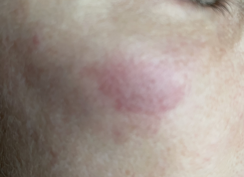
Shortly after completing the trip, the patient noticed a 2.5 cm x 1.5 cm area of non-blanching white surrounded by erythema on the right cheekbone that felt firm to palpation. The patient reported the area to be tender “like a bruise.” The patient kept the area from any further freezing cold exposure. The white lesion to the cheek was presumed to be frostbite, and the face was rewarmed in a warm shower. The white lesion turned purple and was still pliable to touch within 1 hour (Image 1).

There was no bleeding or blistering, but the area began to scab and formed eschar within 12 hours. There was a darker, necrotic-appearing eschar within 24 hours. No bone scan or surgical debridement was felt necessary, and topical wound care was initiated.4 The patient took ibuprofen and a baby aspirin, but thrombolytics were not deemed necessary due to the focal nature of the injury and low likelihood of significant morbidity.1 The palpable defect in the musculature under the necrotic area became more pronounced over time and was most noticeable at 3 weeks, after which it started to resolve.
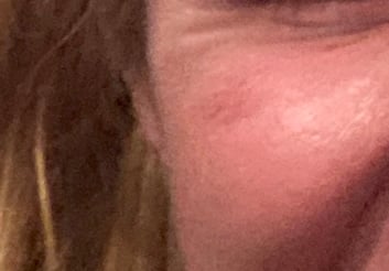
Eschar fell off during the fourth week, revealing tender, friable, erythematous skin underneath and the palpable defect slowly resolving (Image 2).

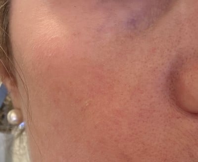
The patient was advised to aggressively avoid cold exposure and keep the area out of the sun. After 5 weeks, the area remained mildly erythematous with a residual palpable defect underneath but was deemed to be improving and was expected to continue to improve. Six months after the cold injury, the skin was nearly normal with some mild residual erythema (Image 3).

Discussion
Immediate or short-term injection necrosis after dermal filler is reported in the literature as a rare but serious side effect that occurs immediately up to several hours after the injection.2 It is usually attributed to direct injection into a vessel or vessel compression due to filler proximity and compression.2 To our knowledge, tissue necrosis 7 weeks after filler has never been reported in the literature.2
In a Finnish study, the most common site of the face and head for frostbite was the ear (58%), followed by the nose (22%), and the cheeks and other parts of the face (20%).9 This is a case of isolated frostbite of the cheek over an area of relatively recent dermal filler when the ears, nose, and the rest of the face had equivocal cold exposure but were spared frostbite injury.
Wilderness Medical Society frostbite guidelines note frostbite can be divided into 4 phases: pre-freeze, freeze-thaw, vascular stasis, and late ischemic.1
- Pre-freeze phase includes cooling, vasoconstriction, and ischemia without ice crystal formation.1
- Freeze-thaw phase includes the formation of ice crystals inside the cells during rapid freezing and outside the cells during a slow freeze.1
- Freezing leads to cellular dehydration, cell membrane lysis, and eventually cell death.1 It can cause local abnormalities in proteins and lipids, as well as shifts in the electrolyte balance of the cell.1
- Thawing leads to reperfusion injury and inflammation.
During vascular stasis, there is alternating constriction and dilation of vessels, leakage from vessels, and coagulopathy in small vessels.1 There is a multifactorial inflammatory response modulated by thromboxane A2, bradykinins, and histamine during the late ischemic phase.1 Ice crystals in and around the tissue cause the initial damage, and disruption to the microcirculation causes cell death.1 Cell damage caused by the ice crystals is made worse by thawing and refreezing.1 The most common anatomic sites of frostbite are the ears, nose, fingers and toes.1,3,8,9 An isolated focal frostbite injury on the cheek that spares the nose and ears is uncommon.
Management
Topical emollients were used in this case initially and as part of ongoing wound care. They are often recommended in wound care to maintain elasticity and to keep the skin moist. While helpful in wound healing, in several studies, topical emollients were not deemed to be significantly protective in preventing initial or further cold injury.5,6,7
Conclusion
While there are other classification systems described in the literature,3 our teams utilize the WMS Classification Description. Based on the WMS description of frostbite classification, this event may have been a focal fourth-degree frostbite injury due to the depth of the palpable tissue defect to the bone. The palpable muscular and tissue defect at 3 weeks was due to the dermal filler under healing but friable skin after the eschar fell off. We believe this apparent focal cold injury could have been augmented by the injected dermal filler elevating the soft tissue off the bone.
TAKE-HOME POINTS
- Frostbite is a preventable condition that occurs when local tissue heat loss exceeds the ability of body perfusion to warm the tissue.1 To our knowledge, this is the first reported case of frostbite likely linked to loss of perfusion capability possibly induced by foreign substance filler cheekbone augmentation.
- The dermal filler elevates the tissue off the bone and occupies space between the tissue and bone.2 As dermal filler facial augmentation becomes more mainstream and this population participates in outdoor cold weather activities, an increased risk of cold injury in thin tissue over dermal filler remains possible.
- Patients undergoing dermal filler augmentation should be vigilant about cold injury when adventuring in extreme cold environments.
Author Contributions
- Sarah Spelsberg performed the direct patient care and most of the writing.
- Andi Sharp assisted with literature review, writing, and editing.
- Sara Filmalter assisted with literature review, writing, and editing.
- Jessica Pelkowski assisted with literature review, writing, and editing.
- Abraham Boxx assisted with literature review, writing, and editing.
References
- McIntosh S, Opacic M, Freer L, et al.Wilderness Medical Society Practice Guidelines for the Prevention and Treatment of Frostbite: 2014 Update. Wilderness Environ Med. 2014; S43-S54.
- Souza Felix Bravo B, Klotz De Almeida Balassiano L, Roos Mariano Da Rocha C, et al. Delayed-type Necrosis after Soft-tissue Augmentation with Hyaluronic Acid. J Clin Aesthet Dermatol. 2015;8(12):42-47.
- Cauchy E, Chetaille E, Marchand V, Marsigny B. Retrospective study of 70 cases of severe frostbite lesions: a proposed new classification scheme. Wilderness Environ Med. 2001;12(4):248-55.
- Banzo J, Martínez Villén G, Abós MD, et al. Congelaciones de manos y pies en un montañero de elite: utilidad de la gammagrafía ósea para predecir el nivel de amputación [Frostbite of the upper and lower limbs in an expert mountain climber: the value of bone scan in the prediction of amputation level]. Rev Esp Med Nucl. 2002;21(5):366-9.
- Lehmuskallio E. Emollients in the prevention of frostbite. Int J Circumpolar Health. 2000;59(2):122-30.
- Lehmuskallio E, Rintamäki H, Anttonen H. Thermal effects of emollients on facial skin in the cold. Acta Derm Venereol. 2000;80(3):203-7.
- Lehmuskallio E. Cold protecting ointments and frostbite. A questionnaire study of 830 conscripts in Finland. Acta Derm Venereol. 1999;79(1):67-70.
- McLeron K, ed. State of Alaska Cold Injury Guidelines. 7th ed. Juneau, AK: Department of Health and Social Services Division of Public Health Section of Community Health and EMS; 2003.
- Lehmuskallio E, Lindholm H, Koskenvuo K, Sarna S, Friberg O, Viljanen A. Frostbite of the face and ears: epidemiological study of risk factors in Finnish conscripts. BMJ. 1995;311(7021):1661-3.


