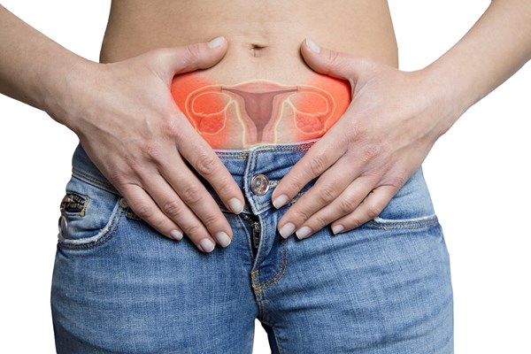
Uterine retroversion is considered a normal variant in an estimated 15% of first-term pregnancies.1
In most cases, retroversion self-corrects around the 10th to 14th week of pregnancy as the gravid uterus ascends from the pelvis into the abdomen. In some cases, the uterus becomes impacted between the sacral promontory and symphysis pubis, and the patient commonly presents with urinary complaints such as acute urinary retention or urinary frequency. Prompt diagnosis in the ED is paramount to reducing obstetric and/or urogynecologic complications. We present a case of a retroverted, incarcerated uterus presenting as acute worsening of urinary retention in a gravid ED patient.
Case Report
The patient is a 32-year-old G2P1 woman with an intrauterine pregnancy at 13 weeks gestation by dates. She presented to the ED complaining of urinary retention. Her symptoms had started about a week earlier and had worsened over the past 12-24 hours. Her symptoms were initially relieved by positional changes (i.e., lying on her side, standing up, sitting on a yoga ball) but then worsened, prompting her visit to the ED. She denied fevers, chills, vaginal bleeding, or other acute systemic symptoms. Her past medical history was relatively non-contributory but included depression, anxiety, insomnia, and undifferentiated connective tissue disease.
The patient’s vital signs were: temperature 36.8 C; blood pressure 120/69; heart rate 89; respiratory rate 16; and oxygen saturation 100% on room air. A physical examination revealed mild abdominal fullness of the lower abdomen as well as a gravid, non-tender uterus. An initial pelvic exam was deferred until a chaperone could be present.
Soon after arrival, a point-of-care ultrasound revealed a single, live intrauterine pregnancy (IUP) with a low-lying uterus, and a large bladder without evidence of hydronephrosis. Additional point-of-care ultrasound imaging was performed for further evaluation of IUP that was grossly normal and consistent with dates. Urine samples were taken to evaluate for asymptomatic bacteriuria of pregnancy.
Given the urinary retention, a Foley catheter was placed with resolution of symptoms. External genitalia examination with a chaperone present revealed a cervix visible at the level of the introitus, raising concern for uterine prolapse causing urethral hypermobility and resultant urinary retention. The diagnosis of retroverted incarcerated uterus was also considered upon review of the literature. Obstetrics was not immediately available for consultation, but the patient was instructed to follow up with family medicine and her midwife, and therefore was felt to be safe for discharge and outpatient follow-up.
The patient followed up with her obstetrician, and while the radiology ultrasound was unrevealing, a physical examination confirmed the diagnosis. The uterus was unable to be manually manipulated in the outpatient setting, though a subsequent trans-rectal reduction under anesthesia was successful.
Discussion
Urinary retention is not an uncommon presenting symptom in ED patients. However, urinary retention in a gravid patient is cause for concern. The differential diagnosis in a gravid patient typically includes: infectious causes such as cystitis or urethritis; neurologic causes like multiple sclerosis; medication effect; and uterine prolapse with urethral hypermobility.2 While it is considered an obstetric emergency, gravid uterine incarceration is rarely included as it is relatively uncommon, occurring in 1 in 3000 pregnancies.1
Uterine incarceration occurs when the uterus becomes stuck between the sacral promontory and the symphysis pubis. This typically occurs between the 14th and 16th week of gestation, and the patient commonly presents with urinary symptoms, such as this patient described.1 The exact mechanism of this process is thought to be from progressive elongation and anterior displacement of the cervix as the gravid uterus expands into the pelvic cavity.3 This causes obstruction of the bladder and urethra, leading to common presenting symptoms such as urinary retention, dysuria, urgency, and paradoxical incontinence. Other common, non-specific presenting symptoms include: abdominal, pelvic, perineal, or back pain; vaginal bleeding; tenesmus; and constipation.4 Identified risk factors for this condition include history of prior incarceration, pelvic inflammation, endometriosis, adhesive disease from abdominal surgeries, and uterine anomalies.4
Due to the highly non-specific nature of presenting symptoms, gravid uterine incarceration can be an easily missed diagnosis. While historically diagnosed clinically, several case reports have noted the ability of imaging studies such as ultrasound and MRI to augment diagnostic potential.4, 5 With the availability of point-of-care ultrasound (POCUS) in many emergency departments, it is important to note that if there is not high clinical suspicion for incarcerated uterus, imaging studies can be easily misinterpreted, leading to a missed diagnosis.
Treatment differs depending on gestational age, though prompt recognition and obstetric consultation are vital to mitigate complications.5
Given the non-specific presenting symptoms, incarceration of a gravid uterus is rarely diagnosed in the ED setting. However, it is a condition with potentially fatal consequences, and the ED clinician should consider it in the differential for the gravid patient presenting with urinary retention.
Teaching Points
- Consider retroverted incarcerated uterus as a cause for acute urinary retention in gravid patients, especially between 14 and 16 weeks gestation.
- MRI is preferred to ultrasound for diagnosis, and transabdominal is preferred to transvaginal ultrasound, though an incarcerated uterus is best diagnosed through history and physical exam.
- Consult OB if there is concern for this diagnosis, as the patient may require urgent reduction, close follow-up, and monitoring for the remainder of the pregnancy.
References
- Sweigart AN, Matteucci MJ. Fever, Sacral Pain, and Pregnancy: An Incarcerated Uterus. West J Emerg Med. 2008;9(4):232-234.
- Serlin DC, Heidelbaugh JJ, Stoffel JT. Urinary Retention in Adults: Evaluation and Initial Management. Am Fam Physician. 2018;98(8):496-503.
- Yang JM, Huang WC. Sonographic Findings in Acute Urinary Retention Secondary to Retroverted Gravid Uterus: Pathophysiology and Preventive Measures. Ultrasound Obstet Gynecol. 2004;23(5):490-495.
- Han C, Wang C, Han L, et al. Incarceration of the Gravid Uterus: A Case Report and Literature Review. BMC Pregnancy Childbirth. 2019;19(1):408. doi:10.1186/s12884-019-2549-3
- Gottschalk EM, Siedentopf JP, Schoenborn I, Gartenschlaeger S, Dudenhausen JW, Henrich W. Prenatal Sonographic and MRI Findings in a Pregnancy Complicated by Uterine Sacculation: Case Report and Review of the Literature. Ultrasound Obstet Gynecol. 2008;32(4):582-586. doi:10.1002/uog.6121


