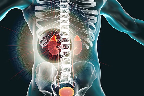
Multiple endocrine neoplasia (MEN) syndromes are rare, inherited disorders in which several endocrine glands develop non-cancerous (benign) or cancerous (malignant) tumors or grow excessively without forming tumors.
There are multiple subtypes of MEN depending on the cluster of abnormal growths. Type 2A involves the thyroid, adrenal or parathyroid glands. Adrenal hemorrhage as the inciting cause is a very rare etiology of abdominal pain, accounting for 0.14%-1.8% in post-mortem analysis,1 but in patients with hemorrhages underlying mass lesions are the most common cause, and pheochromocytoma is the most common identified mass.2 Overall the chief complaint of abdominal pain accounts for 4-5 % of emergency department visits per year, making adrenal hemorrhages a rare cause of a very common complaint.3 We present a case of bilateral adrenal hemorrhages due to pheochromocytomas and MEN2A syndrome.
Case
A 47-year-old female with a medical history significant for type 2 diabetes mellitus, hyperlipidemia, multinodular goiter, hypertension, and congenital left eye blindness presented to the ED because of significant abdominal pain worsening over the past several weeks. Her abdominal pain was poorly localized, and the patient could not identify where her pain began, but only that it is getting significantly worse. Her pain is associated with nausea, vomiting, subjective fevers, fatigue, and weight loss. Social history is significant for cannabinoid use. Past surgical history was significant for laminectomy and peroneal nerve decompression. Patient states that she has not been able to take her blood pressure medications due to persistent vomiting and has been intermittently compliant with her home medications in the recent past.
In the emergency department, her vital signs were significant for tachycardia (122 bpm), and hypertension (176/137). Otherwise she was afebrile, with normal respiratory rate and normal oxygen saturation. Her physical exam was significant for an elevated heart rate with no audible murmurs or additional heart sounds. Lungs were clear to auscultation bilaterally. Her abdominal exam was significant for diffuse abdominal tenderness with both rebound and involuntary guarding. The rest of her physical exam was non-contributory.
Due to the lack of focality on physical exam in conjunction with no signs of impending decompensation, the decision was made to order a full complement of labs to help guide our choice of imaging modality. At this time due to the vague abdominal exam and extended duration of symptoms, we were concerned for many different intra abdominal processes, including pyelonephritis, cystitis, biliary pathology, trauma, bowel obstructions, appendicitis, among others.
Initial laboratory findings were significant for a minimal white blood cell count elevation, 11.8k, with left shift, elevated blood glucose level (308), prerenal azotemia (BUN:Cr - 46:1.64) with an estimated GFR of 37, a lactate of 4.31 without acidosis, and a troponin elevation to 0.196 (negative <0.15). Due to the impaired renal function, the decision was made to get abdominal imaging without an intravenous contrast load. In the meantime the patient received two liters of IV fluids (Ringer’s lactate), Ondansetron 8 mg IV for nausea, and Ketorolac 15 mg for pain. Previous labs were reviewed at the time of presentation and showed a normal GFR.
CT scan was significant for bilateral adrenal gland hemorrhages. Due to the history of subjective fevers and sepsis as a potential cause of adrenal hemorrhage, blood cultures and broad spectrum IV antibiotics (vancomycin and cefepime) were ordered. Endocrinology was also consulted and they recommended IV hydrocortisone 100 mg. At this point the patient was admitted to the ICU for further management, close monitoring and to continue the search for an underlying cause of this presentation. During her admission, additional laboratory findings were diagnostic for pheochromocytoma, including a metanephrine level of >40,000, with a normal level less than 205. Additional findings showed an elevated calcitonin level. The constellation of findings and labs led to the diagnosis of Multiple Endocrine Neoplasia 2A (MEN 2A). Patient was discharged after being medically optimized, and was awaiting outpatient surgical intervention for bilateral hemorrhages/pheochromocytomas. She was admitted to the hospital for a total of 13 days, and spent all 13 of them in the ICU due to the requirement of intensive and difficult to control blood pressure. In the ED we did not treat her blood pressure due to concern for possible progression to hypotension from hemorrhage and possible septic shock. There is no consensus on a goal blood pressure for pheochromocytoma. She was initially treated with Hydralazine, but with minimal efficacy, the patient was transitioned to a titratable Cardene drip. Eventually discharged on a combination of calcium channel blockers, diuretics, and beta blockers. If pheochromocytoma is on the differential, it is important in the ED to remember not to give beta blockers due to the risk of worsening hypertension from unopposed alpha adrenergic stimulation.
Discussion
Adrenal hemorrhage is a rare and potentially life threatening cause of abdominal pain and shock.4 The etiology of adrenal hemorrhage is varied and includes trauma, adrenal lesions, anticoagulation therapy, congenital or acquired bleeding disorders, sepsis due to certain organisms, and pregnancy.4 In our patient due to the lack of trauma, pertinent history, medication uses, the two most likely candidates were an underlying lesion or sepsis. Sepsis due to Neisseria meningitidis is the most common cause of adrenal hemorrhage, with less common organisms H. influenzae, S. pneumoniae, S. aureus, N. gonorrhoeae, P. aeruginosa, and K. oxytoca.5 During her hospital stay, blood cultures were drawn and never grew an identifiable organism. Her markedly elevated metanephrine levels indicated that the cause was more likely to be an adrenal mass.
Adrenal hemorrhage management is complicated as it must address two different problems - adrenal crisis often with accompanying shock, and hemorrhage from adrenal glands which can cause hypovolemic shock and may necessitate adrenal artery angioembolization.6,7 Hypovolemic shock is primarily managed by surgical or interventional radiology intervention. While no definite management guidelines exist, consensus states that early IV hydrocortisone 100 mg prevents deterioration and shock, and should be started if IV fluids resuscitation does not adequately support blood pressure.8
Pheochromocytomas are catecholamine secreting tumors that arise from the medulla of the adrenal glands.9 They are rare, occurring in less than 0.2 percent of patients with hypertension with hypertension being one of the most common presenting findings of pheochromocytomas.10 The classic presentation of pheochromocytoma is headache, hypertension, and sweating occurring paroxysmally over several months to years. Our patient on presentation denied having any headaches or sweats but was noted to be hypertensive. Given her history of hypertension and medication non-compliance we did not initially associate her elevated blood pressure with her acute complaints.
In patients in whom one suspects a pheochromocytoma, the most commonly used and accurate tests are both serum and urine metanephrine screens. Metanephrines are metabolites of catecholamines, which are produced by the rapidly reproducing chromaffin cells of pheochromocytoma tumors.11 Management of pheochromocytomas includes a combination of surgical intervention and chemotherapy - a combination therapy of vincristine, cyclophosphamide, and dacarbazine. For the emergency clinician, controlling blood pressure is of vital importance, especially before any surgical intervention can take place. Initial management involves alpha adrenoceptor blockade, before beginning conventional beta adrenergic blockade therapy.12
As part of syndromes, pheochromocytomas occur in Multiple Endocrine Neoplasia Type 2, both 2A and 2B. Unlike pheochromocytomas in regular patients, pheochromocytomas that arise as part of a genetic syndrome do not generate the same symptoms as in non-syndromic presentations. Half of patients with pheochromocytomas as part of MEN 2 do not have symptoms, and only one third of patients have hypertension.13 A similar finding is present in patients with Von Hippel Lindau disease, with 35% of VHL pheochromocytoma patients being asymptomatic.14
Multiple endocrine neoplasia type 2 (MEN2) is a rare disorder involving the development of medullary thyroid carcinoma, unilateral or bilateral pheochromocytomas, and proliferation of other endocrine tissues within the same individual.15
Abdominal pain, as a presenting complaint to the ED, will continue to be one of the more complicated syndromes we face, as it can be caused by multiple different disease processes involving multiple different organ systems. While rare, the presentation of an endocrine emergency, more specifically adrenal hemorrhage, as a consequence of MEN2 pheochromocytomas is potentially life threatening and requires a multidisciplinary approach to management. Her presentation of diffuse abdominal pain, in the setting of adrenal hemorrhage while hypertensive is not common, and should prompt more extensive evaluation. After admission, and medical optimization in the ICU patient was safely discharged to outpatient surgical follow up. Two weeks later she underwent surgical removal of the tumors and hematomas, and is currently doing well.
References
- Rao RH. Bilateral massive adrenal hemorrhage. Med Clin North Am 79: 107–29, 1995.
- Marti, J.L., Millet, J., Sosa, J.A. et al. Spontaneous Adrenal Hemorrhage with Associated Masses: Etiology and Management in 6 Cases and a Review of 133 Reported Cases. World J Surg 36, 75–82 (2012). https://doi.org/10.1007/s00268-011-1338-6
- Powers RD, Guertler AT. Abdominal pain in the ED: stability and change over 20 years. Am J Emerg Med. 1995 May;13(3):301-3. doi: 10.1016/0735-6757(95)90204-X. PMID: 7755822
- Karwacka IM, Obołończyk Ł, Sworczak K. Adrenal hemorrhage: A single center experience and literature review. Adv Clin Exp Med. 2018 May;27(5):681-687. doi: 10.17219/acem/68897. PMID: 29616752.
- Guarner, J., Paddock, C., Bartlett, J. et al. Adrenal gland hemorrhage in patients with fatal bacterial infections. Mod Pathol 21, 1113–1120 (2008). https://doi.org/10.1038/modpathol.2008.98
- Logaraj A, Tsang VH, Kabir S, Ip JC. Adrenal crisis secondary to bilateral adrenal haemorrhage after hemicolectomy. Endocrinol Diabetes Metab Case Rep. 2016;2016:16-0048. doi:10.1530/EDM-16-0048
- Ali A, Singh G, Balasubramanian SP. Acute non-traumatic adrenal haemorrhage-management, pathology and clinical outcomes. Gland Surg. 2018;7(5):428-432. doi:10.21037/gs.2018.07.04
- Bashari WA, Myint YMM, Win ML, Oyibo SO. Adrenal Insufficiency Secondary to Bilateral Adrenal Hemorrhage: A Case Report. Cureus. 2020;12(6):e8596. Published 2020 Jun 13. doi:10.7759/cureus.8596
- Young, W. F., Jr. (2020, August 31). Clinical presentation and diagnosis of pheochromocytoma. Retrieved November 17, 2020, from https://www.uptodate.com/contents/clinical-presentation-and-diagnosis-of-pheochromocytoma
- Stein PP, Black HR. A simplified diagnostic approach to pheochromocytoma. A review of the literature and report of one institution's experience. Medicine (Baltimore). 1991 Jan;70(1):46-66. doi: 10.1097/00005792-199101000-00004. PMID: 1988766.
- Eisenhofer G, Prejbisz A, Peitzsch M, Pamporaki C, Masjkur J, Rogowski-Lehmann N, Langton K, Tsourdi E, Pęczkowska M, Fliedner S, Deutschbein T, Megerle F, Timmers HJLM, Sinnott R, Beuschlein F, Fassnacht M, Januszewicz A, Lenders JWM. Biochemical Diagnosis of Chromaffin Cell Tumors in Patients at High and Low Risk of Disease: Plasma versus Urinary Free or Deconjugated O-Methylated Catecholamine Metabolites. Clin Chem. 2018 Nov;64(11):1646-1656. doi: 10.1373/clinchem.2018.291369. Epub 2018 Aug 10. PMID: 30097498.
- Nölting S, Ullrich M, Pietzsch J, et al. Current Management of Pheochromocytoma/Paraganglioma: A Guide for the Practicing Clinician in the Era of Precision Medicine. Cancers (Basel). 2019;11(10):1505. Published 2019 Oct 8. doi:10.3390/cancers11101505
- Pomares FJ, Cañas R, Rodriguez JM, Hernandez AM, Parrilla P, Tebar FJ. Differences between sporadic and multiple endocrine neoplasia type 2A phaeochromocytoma. Clin Endocrinol (Oxf). 1998 Feb;48(2):195-200. doi: 10.1046/j.1365-2265.1998.3751208.x. PMID: 9579232.
- Walther MM, Reiter R, Keiser HR, Choyke PL, Venzon D, Hurley K, Gnarra JR, Reynolds JC, Glenn GM, Zbar B, Linehan WM. Clinical and genetic characterization of pheochromocytoma in von Hippel-Lindau families: comparison with sporadic pheochromocytoma gives insight into natural history of pheochromocytoma. J Urol. 1999 Sep;162(3 Pt 1):659-64. doi: 10.1097/00005392-199909010-00004. PMID: 10458336.
- Marini F, Falchetti A, Del Monte F, et al. Multiple endocrine neoplasia type 2. Orphanet J Rare Dis. 2006;1:45. Published 2006 Nov 14. doi:10.1186/1750-1172-1-45.


