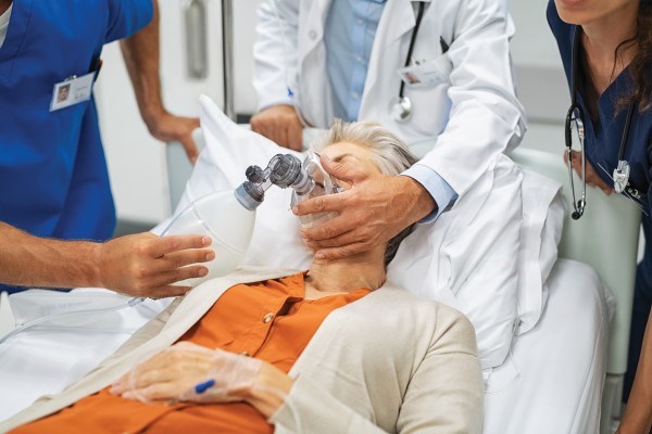
A 76-year-old female presented to the ED via EMS after a friend found her at home with a decreased level of consciousness and dyspnea.
She was discovered by EMS to be markedly hypoxic, bradycardic, hypotensive, and hypothermic. She was discharged from another hospital approximately one month prior for management of a small subarachnoid hemorrhage. During her workup, her TSH was found to be 70.2 mIU/mL.
Pathophysiology and Causes
In healthy humans, the hypothalamus produces thyrotropin-releasing hormone (TRH), which stimulates the anterior pituitary to release thyroid-stimulating hormone (TSH). This hormone subsequently stimulates the thyroid gland to produce thyroxine in its two forms: T4 and T3. The receptors for thyroxine are found in almost every cell in the body and enable it to regulate homeostasis.
When this system is disrupted through certain pathological conditions, the ability for the body to maintain homeostasis is impaired. An insult to this regulatory system that results in a deficiency of thyroxine is called hypothyroidism, while an excess is called hyperthyroidism. Either condition results in cardiac, neurological, respiratory, gastrointestinal, hematologic, renal, and electrolyte abnormalities.1
In this case, we will discuss hypothyroidism. In short, hypothyroidism slows down metabolism in the human body and impairs its ability to respond to adverse conditions that would normally require the body to compensate in order to maintain homeostasis.
Clinical Presentation and Diagnosis
Severe hypothyroidism is commonly referred to as “myxedema coma.” However, patients with this condition are often not comatose and do not have edema. Rather, they are most notably recognized by declining mental status along with findings of an inciting event that led to dysregulation.1 Inciting events can be anything from infection, drug effects, ischemia, metabolic abnormalities, hypoxia, cold exposure, or even forgetting to take prescribed supplemental thyroxine.
Consequently, recognizing common manifestations of severe hypothyroidism is key to forming the differential diagnosis.
Cardiac manifestations are most easily seen on an electrocardiogram (ECG). Some common findings include: bradycardia, flattened T-waves, bundle branch blocks, and complete heart block.
Neurologically, the most common finding is an altered mental status.
Respiratory symptoms include hypoxia and hypercapnia, which can be a result of decreased respiratory drive.
Gastrointestinal symptoms are vague, but include nausea, vomiting, abdominal pain, constipation, ileus, ascites, and even anasarca.
Renal function is decreased, and hyponatremia is a common electrolyte abnormality.
Lastly, hematologic manifestations include an increased risk for bleeding due to an acquired deficiency of many clotting factors.2
It can be difficult to recognize all of these symptoms together when a patient presents to the emergency room, as there are often multiple pathological processes present at the time of diagnosis. However, the typical patient profile is as follows: female, elderly, known hypothyroidism or status post-thyroidectomy, hypothermic, altered mentation, refractory hypotension, hypoxia with hypercapnia, bradycardia, edema, acute precipitating illness, drug toxicity, and hyponatremia.2 Essentially, a patient will be slow, cold, and in extreme distress.
The diagnosis of severe hypothyroidism can be achieved with a single TSH along with a free T4 (FT4). In primary hypothyroidism, TSH will be increased and FT4 will be decreased. Central hypothyroidism will have a decreased or normal TSH with a decreased FT4.
Regardless of the cause, severe hypothyroidism has a high mortality risk (29 percent), so identification and prompt treatment is of paramount importance in the critically ill patient.3
Treatment
- Treat any underlying issue or inciting event.
- Rapid replacement of thyroxine; initial IV load of 200-400mcg.
- Empiric stress-dose steroids are recommended. A single dose of 100mg IV hydrocortisone is appropriate.
- Hypotension may respond to crystalloid, but pressors may be required.
- Passive or active rewarming.
- Treat any hyponatremia with use of hypertonic saline only with severe cases of altered mental status or seizures.
Case Conclusion
Initially, the differential diagnosis in this case was broad. Treatment was empiric as an underlying inciting event was suspected for the patient’s presentation. Atropine, calcium chloride, and glucagon were administered. A dopamine infusion was started, and broad-spectrum antibiotics were initiated. As time passed, the patient’s respiratory distress worsened, and she was intubated utilizing ketamine and rocuronium. However, as laboratory tests began to result and additional history was obtained on family arrival, the diagnosis of severe hypothyroidism became a likely possibility. According to the patient’s family, the patient had unintentionally missed over one month of her supplemental levothyroxine. Thus in addition to the initial resuscitation, stress-dose steroids and IV levothyroxine were administered, and the patient was admitted to the intensive care unit for further management and care.
Clinical Pearls
- Patients can present with unstable vitals, including hypothermia, hypotension, bradycardia, hypoxia, and bradypnea.
- Patients with severe hypothyroidism do not always have edema and are not always comatose, as implied by the term “myxedema coma.”
- If you suspect severe hypothyroidism, obtain a TSH with free T4; these are relatively quick and diagnostic.
- Critical steps include a bolus of IV levothyroxine, 200-400mcg, and a stress-dose steroid bolus of hydrocortisone 100mg IV.
- Electrocardiogram changes are nonspecific, but sinus bradycardia and nonspecific ST-segment changes are common.
- Treat any underlying condition, as this was likely the inciting event that preceded the patient’s presentation and led to decompensation.
References
- Elshimy G, Chippa V, Correa R. Myxedema - StatPearls - NCBI Bookshelf. Myxedema. https://www.ncbi.nlm.nih.gov/books/NBK545193/. Published August 22, 2022. Accessed January 22, 2023.
- Walls RM, Hockberger RS, Gausche-Hill M, et al., eds. Rosen's Emergency Medicine: Concepts and Clinical Practice. Vol 2. 10th ed. Philadelphia, PA: Elsevier Health Sciences; 2023.
- Talha A, Houssam B, Brahim H. Myxedema Coma: A Review. European Journal of Medical and Health Sciences. 2020;2(3). doi:10.24018/ejmed.2020.2.3.349


