


A patient's insistence that something was living under her skin led to yet another application of a popular diagnostic tool.
Case
A 20-year-old female patient with no significant past medical history presented to our emergency department (ED) with itchy scalp lesions after returning from a honeymoon to Belize. Over the past 3 weeks, she had presented to the clinic several times, each time being treated for “bug bites” with topical steroid cream. At her most recent clinic visit, the patient described feeling strange sensations like wriggling under her skin. She was provided a psychiatry referral out of concern for delusional parasitosis.
The morning prior to presentation, the patient’s partner cleaned her scalp and noted something moving under her skin (Figure B). Bedside ultrasound performed in the ED demonstrated two large foreign bodies within the scalp, with internal movement (Figure A and video). We discussed treatment options with the patient, including watchful waiting, occlusion of the breathing pore, or procedural excision. After discussing the risks and benefits, the patient elected for bedside excision, which was completed using ultrasound guidance (see below) (Figure C). The patient tolerated the excision well. Specimens were sent to the pathology lab for analysis and were consistent with Dermatobis hominis (human bot fly).
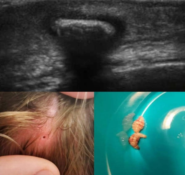
Figure 1: Cutaneous myiasis of the scalp. A. Still of the ultrasound image that was used to identify one of the larvae (arrow) at the time of initial evaluation in the emergency department. B. Appearance of the patient’s scalp with prominent breathing pore (arrow). C. Two live, intact bot fly larvae (arrow) post excision.
Video: BotflyScalpMirrielees.mp4
Cutaneous myiasis of the scalp, ultrasound video clip. This video demonstrates the bot fly larva (arrow) embedded in the patient’s scalp. The wriggling movement in the video narration represents the larva’s spicules, which it uses to grip the host skin in order to resist expulsion.
Procedure Figure: Approach to bedside excision. Figure above demonstrates the larva embedded in the skin with an overlying pore (dark circle). Linear ultrasound probe was used to identify the long axis of the larva and a small skin incision (dashed red line) was made.

About the Bot Fly
The human bot fly (D. hominis) is endemic to Central and South America, transmitted primarily via mosquito bite,1 leading to cutaneous myiasis. There is a distinct species of bot fly (Cordylobia) that is endemic to the African tropics. Therefore, patients returning from an endemic area may have appropriate concern when presenting to your ED with feelings of something moving under their skin, and you should have a higher index of suspicion for a parasitic etiology in such patients.
The bot fly life cycle begins with the female laying eggs in a mosquito or other vector insect. The eggs are harbored in the insect’s abdomen and released into a host during feeding. After seeding of the host, self-expulsion of the larva typically occurs after 5-10 weeks. Disclosing this time frame to patients is important when discussing expectant management.
There are many different species of bot fly that primarily affect other mammals, and rarely, if ever, transmit to humans.2
About the Procedure
Prior to incision, the site of each larva was treated with lidocaine-tetracaine-epinephrine (LET) gel for topical anesthesia, with a secondary benefit of stunning the larva (larvae that are not adequately weakened either pharmacologically or via asphyxiation are likely to resist removal via use of their spicules, see discussion below). A linear probe was used to determine the long axis of the larva and optimize the small (less than one cm) incision) made to deliver each larva. Once the incision was made, gentle pressure was applied, and each larva was expelled quickly. The wounds were irrigated with sterile saline and left open to heal by secondary intention with plan for PO antibiotics on discharge.
Management Options to Offer the Patient Presenting with Cutaneous Myiasis
Conventional teaching for management of cutaneous larvae has generally been to asphyxiate the larvae. Asphyxiation encourages the larvae to leave the skin voluntarily or weakens it to facilitate manual removal by squeezing the larvae out.
Attempting to remove the larvae without prior asphyxiation or stunning is not recommended, as the larvae unfortunately resist removal by digging into host skin with their spicules (see ultrasound video to observe movement of the larva spicules in our case patient).3
Options for asphyxiation include nail polish or petroleum jelly, both of which are intended to weaken the larvae for extraction. An additional option characterized in the literature is applying bacon,4 which reportedly encourages the larvae to leave the skin either through asphyxiation and/or attraction to the bacon or bacon grease itself; some patients may attempt this as a home remedy.
There is also a case report describing the use of a commercial venom extractor.5 There have been prior case reports 6,7 utilizing ultrasound for diagnosis and management, including those describing bedside excision in the ED.8,9
Possible complications include cellulitis, especially if there is any concern that the larva may have been transected or otherwise incompletely removed and dies within the skin, therefore In this case, treatment with a course of antibiotics targeting skin and soft tissue infections is very reasonable.
Conclusion
Cutaneous larvae is an uncommon ED finding in the United States. Frequently patients who describe worms or insects under their skin are not found to have active parasitic infestation; their symptoms may instead be attributable to a wide variety of causes including dry skin, eczema, infection, allergic reaction, or much less commonly, psychiatric illness.
For patients with relevant travel history or clinical exam findings, soft tissue point of care ultrasound can assist in evaluation for active parasitosis along with other etiologies such as fluid collection or foreign body.
In the ED, ultrasound can aid in diagnosis and provide procedural guidance for management of a variety of skin and soft tissue infections. Ultrasound may also have a place as a routine study to complete during the physical exam for these patients.
This case serves as a reminder to complete appropriate evaluation in any patient expressing concern for a parasite with correlating exam findings. When patients present with either nonspecific or suspect (“worms under my skin”) signs or symptoms, bedside ultrasound can offer a very effective and rewarding approach to evaluating skin and soft tissue complaints, especially when clinical gestalt suggests there may be more than meets the eye.
References
- Mattison N, Cooper JS. Botfly. [Updated 2021 Jul 18]. In: StatPearls [Internet]. Treasure Island (FL): StatPearls Publishing; 2021 Jan-. Available from: https://www.ncbi.nlm.nih.gov/books/NBK559223/
- Maier H, Hönigsmann H. Furuncular myiasis caused by Dermatobia hominis, the human botfly. J Am Acad Dermatol. 2004 Feb;50(2 Suppl):S26-30
- McGarry JW. Tropical myiases: neglected and well travelled. Lancet Infect Dis. 2014 Aug;14(8):672-674.
- Brewer TF, Wilson ME, Gonzalez E, Felsenstein D. Bacon therapy and furuncular myiasis. JAMA. 1993 Nov 3;270(17):2087-8. PMID: 8411575.
- Boggild AK, Keystone JS, Kain KC. Furuncular myiasis: a simple and rapid method for extraction of intact Dermatobia hominis larvae. Clin Infect Dis. 2002 Aug 1;35(3):336-8.
- Bovino P, Cole J, Scheatzle M. Ultrasound Visualization of Atypical Abscess Ultimately Containing Bot Fly Larva. J Emerg Med. 2016 Aug;51(2):144-6.
- Schechter E, Lazar J, Nix ME, Mallon WK, Moore CL. Identification of subcutaneous myiasis using bedside emergency physician performed ultrasound. J Emerg Med. 2011 Jan;40(1):e1-3.
- Jones CH, Leon M, Auerbach J, Portillo-Romero J. Ultrasound Detection of Human Botfly Myiasis of the Scalp: A Case Report. Cureus. 2020 Dec 4;12(12):e11905.
- Zamaray M, Hekker TAM, Schuurs TC. Man With Gnawing Sensation Under His Scalp. Ann Emerg Med. 2020 Sep;76(3):363-378.