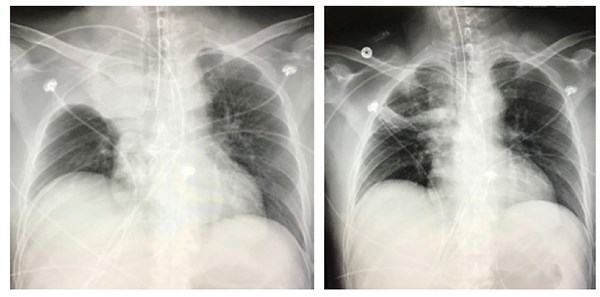
Up to 5% of the population has a right superior lobe bronchus arising directly from the trachea proximal to the carina. It can cause complications with airway management if unrecognized, and can be the root cause of emphysema, atelectasis, hemoptysis, and recurrent pneumonia.
Chief Complaint
Altered mental status and abnormal lung sounds after intubation
History of Present Illness
A 47-year-old-male with a past medical history of chronic alcoholism and possible seizure disorder presented to the ED via ambulance for a grand mal seizure witnessed by a bystander. It was reported that he had been having multiple episodes of seizures preceding this event. While in the ED, the patient continued to be altered and suffered multiple subsequent seizures. The patient was intubated for airway protection. A chest radiograph was taken before and after intubation. The patient had normal lung sounds before intubation and absent lung sounds in the right upper lobe afterward.
Initial Physical Exam
VITALS: BP: 116/75, Pulse: 104, Temp: 101, RR: 16, SpO2: 100% on room air
GENERAL: Patient was nonresponsive and post-ictal. No distress.
NEURO: Altered, unresponsive, nonverbal with minimal moans and eye opening to noxious stimuli, withdrawal noted in all extremities.
RESPIRATORY: Breath sounds clear and equal bilaterally, no respiratory distress.
Physical Exam After Intubation
VITALS: BP: 119/76, Pulse: 105, Temp: 98.3, RR: 16, SpO2: 100% on 100 percent FiO2
GENERAL: Patient intubated and sedated
RESPIRATORY: Absent breath sounds right upper lung
Physical Exam After ET Tube Retracted 2 cm
RESPIRATORY: Clear and equal lungs sounds throughout
Pertinent Radiology
Initial CXR showed no acute disease. Post-intubation chest radiograph demonstrated right upper lobe consolidation with volume loss, which was not present on the initial film. Tip of the endotracheal tube was 10 mm above the carina. Chest radiograph after endotracheal tube retracted 2 cm showed partial resolution of the right upper lobe consolidation.
- Aspiration or anatomical abnormality such as tracheal bronchus are the key etiologies behind this post-intubation radiographic finding.
- Look for specific lung sounds and physical exam findings with this abnormal CXR, including increased fremitus on the side with consolidation, dullness to percussion, absent breath sounds or crackles, or increased vocal resonance.
Case Discussion
It is generally important to repeat radiographs after certain procedures, especially with intubation. In this case, the patient was found to have an anatomical variant called a tracheal bronchus. In 0.1-5% of the population there is a right superior lobe bronchus arising directly from the trachea proximal to the carina.
It can have multiple variations and, although usually asymptomatic, it can be the root cause of emphysema, atelectasis, hemoptysis, and persistent or recurrent pneumonia.
Computed tomography is the best modality for assessing the anatomy and allows direct visualization and orientation of the anomalous bronchus.
It is important for physicians who perform advanced airway management to be knowledgeable about this anatomical variant, so that prompt recognition can prevent delays in diagnosis and management in the acute care setting.
Pearls
- Significant change in lung sounds after intubation can alert us to complications, and if lungs sound are abnormal in the right upper lobe it could be caused by a tracheal bronchus.
- Knowledge of anatomical variations and complications of procedures can allow for quick identification, management, and improved outcomes.
References
1. Weerakkody Y, et al. Tracheal Bronchus. Radiopaedia. 2007. Accessed 2018.
2. Bakir B, Terzibasioglu E. Images in Clinical Medicine: Tracheal Bronchus. N Engl J Med. 2007;357:1744.


