Ruptured thoracic aortic aneurysms are almost uniformly fatal. What can you do to provide the early detection and treatment that can offer hope of survival?
Case
A 98-year-old woman with past medical history of hypertension, chronic kidney disease stage III, hyperlipidemia, congestive heart failure, and coronary artery disease status post two-vessel coronary artery bypass graft in 2001 presented to a high-volume, suburban emergency department in Florida by EMS with complaint of left-sided flank pain at 6 am.
Her vitals were BP 110/59 mmHg, HR 87 bpm, RR 18 bpm and temperature 36.6°C during transport and on arrival. The patient was alert and oriented on initial exam but in moderate discomfort. History was initially limited due to a language barrier and patient distress. The patient reported upper abdominal pain and left flank pain that started the previous night. She became acutely altered in the exam room with a left-sided gaze deviation. Her vitals remained unchanged and her altered mentation resolved after a period of 1-2 minutes. She was rushed to CT for emergent neurologic imaging as well as imaging of the chest and abdomen.
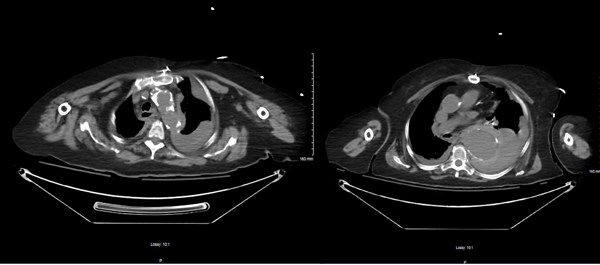
Non-contrast CT of the abdomen and pelvis showed a ruptured thoracic aortic aneurysm [Figure 1] with hemorrhage into the mediastinum and throughout the left pleural cavity with two pseudoaneurysms of the descending thoracic aorta, the largest and most distal of which extended over 6 cm in length and 5.6 cm in depth.
The family was informed of her diagnosis and prognosis and were at bedside. The patient was given comfort measures per family wishes. The patient expired shortly thereafter with PEA arrest.
Total time from patient arrival to expiration was 30 minutes.
Discussion
Ruptured thoracic aortic aneurysms are almost uniformly fatal, with a mortality rate of 97-100%.1 Early detection and intervention are essential to curbing this mortality rate. Risk factors for this patient included advanced age, hypertension and atherosclerosis.2,3
This case demonstrates a rare visualization of an actively rupturing thoracic aneurysm and a non-classical exam presentation.
References
1. Johannson G, Markström U, Swedenborg J. Ruptured thoracic aortic aneurysms: a study of incidence and mortality rates. J Vasc Surg. 1996;21(6):985-988.
2. Forsdahl SH, Singh K, Solberg S, Jacobsen BK. Risk Factors for Abdominal Aortic Aneurysms. Circulation. 2009;119(16):2202-2208.
3. Coady MA, Rizzo JA, Goldstein LJ, Elefteriades JA. Natural history, pathogenesis, and etiology of thoracic aortic aneurysms and dissections. Cardiol Clin. 1999;17(4):615-635.


