This case represents a classic progression of a rarely diagnosed disease and showcases the role of emergency physicians in ensuring patients with rare pathology receive appropriate care and diagnoses during evolving symptomatology.
Case Presentation
A 51-year-old male with past medical history of tobacco use, hyperlipidemia, and bipolar disorder presented to the emergency department for eye pain, eye drainage, and sore throat 11 days after being diagnosed with COVID-19. Initial symptoms of COVID-19 were mild and included fatigue, headache, and non-productive cough. However, 6 days after positive PCR testing, he noticed worsening bilateral conjunctival injection, yellow eye drainage, sore throat, dysuria, and decreased appetite. He did not report fevers, chest pain, shortness of breath, myalgias, arthralgias, or GI complaints. The patient was not vaccinated against COVID-19.
Initial workup showed slight elevations of AST and ALT, significantly elevated CRP of 15.11mg/dL, proteinuria, and urobilinogen. Chest X-ray was unremarkable. Exam was notable for severely injected conjunctiva bilaterally with thick yellow discharge. Skin exam showed multiple circular erythematous macules to the left anterior leg. Oral exam showed few aphthous ulcers and was negative for erythema or tonsillar exudates. He was well-appearing with tachycardia that improved with 1L of crystalloid fluids. Symptoms were felt to be due to viral conjunctivitis, but given eye discharge he was prescribed polymyxin B/trimethoprim eye drops for superimposed bacterial infection. He was discharged and instructed to follow-up with his PCP and ophthalmology.
The patient returned to the ED the following day due to worsening symptoms. Sore throat had progressed significantly, and he now reported hoarseness, increased oral lesions, and pain that limited his ability to eat, swallow, and speak. Bilateral eye pain, swelling and discharge continued to worsen despite treatment with new bilateral subconjunctival hemorrhages. He reported spreading of lower extremity rash to involve bilateral legs and upper extremities. On the day of the second ED visit, he noticed dysuria and a small amount of blood from his urethra after urination. The patient denied other symptoms including myalgias or arthralgias. He had been treated with lamotrigine for bipolar disorder for several years and denied any new medications. He denied a history of STIs, UTIs, or being currently sexually active.
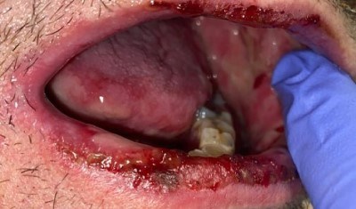
On evaluation, the patient was in obvious discomfort with speaking and swallowing secretions. Oral exam showed diffuse aphthous ulcerations of the mucosa, swelling, and tenderness to palpation (Fig. 1-3). Eye exam was notable for bilateral subconjunctival hemorrhages, yellow discharge, photophobia, and periorbital erythema and swelling (Fig. 4). Genital exam showed thin yellow discharge at the urethral meatus with tenderness to palpation of distal penis. Circular, nonpruritic macules were present over the bilateral anterior legs (Fig. 5).
Figures 1-3. Oral lesions in a patient with Behcet's disease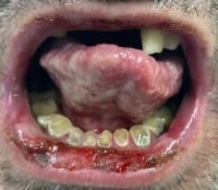
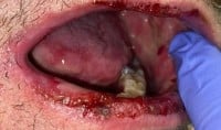
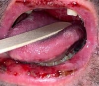
Figure 4. Occular findings in a patient with Behcet's disease
Figure 5. Dermatologic findings in a patient with Behcet's disease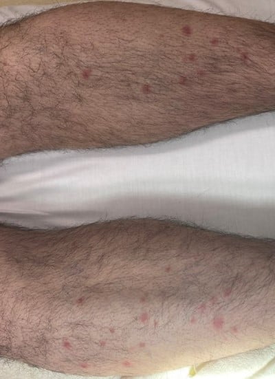
Work-up was notable for AST of 90U/L, ALT of 72U/L, ESR of 38mm/h, CRP 18.44mg/dL, fibrinogen of 745mg/dL, UA showing 2+ protein, and 11-20 WBCs and RBCs. Gonorrhea and chlamydia testing was negative. The patient was given ketorolac, morphine, and oral viscous lidocaine for pain.
Background
Behcet’s disease (BD) is a rare, multisystem inflammatory disease characterized by vasculitis of all vessel sizes. The exact cause of and underlying pathophysiology of BD is unclear, although there are established risk factors including genetic predisposition, male sex, immunological triggers including viral infections or vaccinations, age, and ethnicity. While it is thought to be autoinflammatory in nature, there is no evidence that specific autoantibodies are involved in the development of BD.1-3 Incidence and prevalence vary widely across geographical areas, with an estimated incidence of 0.58 per 100,000 in Middle Eastern and Japanese populations, and incidence of 0.24 per 100,000 in European populations. Studies in Western populations are limited but show an approximate incidence of 0.38 per 100,000 in the Midwest United States.6 Diagnosis of BD is clinical, based on the presence of classic symptoms and supported by a relapsing and remitting course of disease. Multiple diagnostic criteria have been developed with the most established being the International Criteria for Behcet’s Disease which uses a scoring system based on symptoms of oral aphthosis, vascular lesions (DVT, LVT, arterial thrombus, superficial phlebitis and aneurysm), genital lesions, inflammatory eye disease (anterior uveitis, posterior uveitis and retinal vasculitis), rash, and a positive pathergy test.4
More serious complications include bowel perforation from GI ulcerations, ruptured coronary, pulmonary or peripheral aneurysms, pulmonary embolism, stroke, dural venous thrombosis, meningoencephalitis, and blindness. Due to the rarity of BD, multisystem involvement, and relapsing and remitting symptoms, diagnosis is clinically challenging. Patients on average have symptoms for 5.3 years and meet diagnostic criteria for 1.1 years before an official diagnosis is made.
Timely diagnosis and treatment with appropriate, symptom-based therapy is essential in reducing patient morbidity and mortality along the disease course.2 First line treatment is colchicine +/- apremilast and may include oral, topical and ophthalmologic steroids. Severe or refractory symptoms require treatment with azathioprine, interferon-alfa, cyclosporine, methotrexate, and TNF-alpha inhibitors.3
Differential Diagnosis
The differential diagnoses for a patient that presents with oral ulcers, rash, ocular and urinary symptoms are broad and present a diagnostic challenge. Differential diagnoses including HSV, Steven-Johnson syndrome (SJS), reactive arthritis, lupus, measles and in the setting of COVID-19 infection, multisystem inflammatory syndrome must be considered. While rare, it is important to consider the various forms of vasculitis including BD, polyarteritis nodosa, and microscopic polyangiitis depending on the patient's age and demographics. In our patient, history of treatment with lamotrigine increased suspicion of SJS; however, the clinical presentation and symptoms were not typical. Multisystem involvement and recent viral infection helped to narrow our differential diagnosis and the primary concerns were for reactive arthritis and MSIS.
Diagnosis and Treatment
Infectious disease was consulted in the emergency department for known COVID-19 and suspected MSIS as at the time of patient’s presentation, treatment options were rapidly evolving. In the setting of oral aphthous ulcerations, GU symptoms, anterior uveitis/conjunctivitis, and rash following viral infection in a middle-aged male, BD was considered the most likely diagnosis. Using the International criteria for Behcet’s disease, the patient met the criteria score of >4 for the following symptoms: oral aphthous ulcers (2pts), ocular manifestation (2pts), rash.1 Although the patient was complaining of penile pain, no genital ulcerations were noted at the time of evaluation.
Given the severity of symptoms and inability to tolerate oral hydration, the patient was admitted to the hospital for initiation of treatment and supportive therapy. He was given a 3 g loading dose of colchicine, followed by 0.6 mg TID and started on 5mg prednisone daily. During hospital stay he had improvement in symptoms and was discharged to home on colchicine and prednisone with ophthalmology and rheumatology follow-up. Steroids were tapered after one month and after several months without symptoms colchicine was tapered as well.
Discussion
Behcet’s disease is a rare vasculitis that affects large, medium, and small vessels leading to multisystem involvement and a wide variety of symptoms. It is a diagnosis that often requires multiple health care visits for identification and requires appropriate treatment to prevent significant long-term morbidity and increased risk of mortality.
This case demonstrates a classic presentation of BD diagnosed in the ED after 2 visits for worsening oral ulcerations, anterior uveitis, rash, and genital pain in the setting of a recent infection. While long-term management is outside of the scope of the emergency physician, this case demonstrates the importance of identifying abnormal pathology or clinical courses requiring subspecialist consultation and hospitalization. Following appropriate diagnosis and treatment, our patient recovered without long-term effects.
Take-Home Points
- Behcet’s disease is a rare condition that may go undiagnosed for years and carries a high risk of morbidity and mortality.
- Patients with Behcet’s disease most often present with oral aphthous ulcers and ocular disease early in disease course and may have additional organ involvement including rash, genital ulcers, arthritis, and vascular pathology.
- Behce’ts is most often diagnosed in the 2nd-4th decades of life but may occur at any time and often have a preceding immunological trigger including viral infection or recent vaccination.
- Behcet’s disease can lead to significant complications including blindness, aneurysms, bowel perforation, or meningoencephalitis, and patients may require hospitalization for initiation of treatment or stabilization. Prompt diagnosis and treatment is essential in preventing long term sequelae.
References
- National Organization for Rare Disorders. Behçet’s syndrome - symptoms, causes, treatment. May 17, 2023.
- Alpsoy E, Donmez L, Onder M, et al. Clinical features and natural course of Behçet's disease in 661 cases: a multicentre study. Br J Dermatol. 2007;157(5):901-906.
- Adil A, Goyal A, Quint JM. Behcet Disease. [Updated 2023 Feb 22]. In: StatPearls [Internet]. Treasure Island (FL): StatPearls Publishing; 2023.
- Davatchi F. Diagnosis/Classification Criteria for Behcet's Disease. Patholog Res Int. 2012;2012:607921.
- Al-Araji A, Kidd DP. Neuro-Behçet's disease: epidemiology, clinical characteristics, and management. Lancet Neurol.2009;8(2):192-204.
- Calamia KT, Wilson FC, Icen M, Crowson CS, Gabriel SE, Kremers HM. Epidemiology and clinical characteristics of Behçet's disease in the US: a population-based study. Arthritis Rheum.2009;61(5):600-604.


