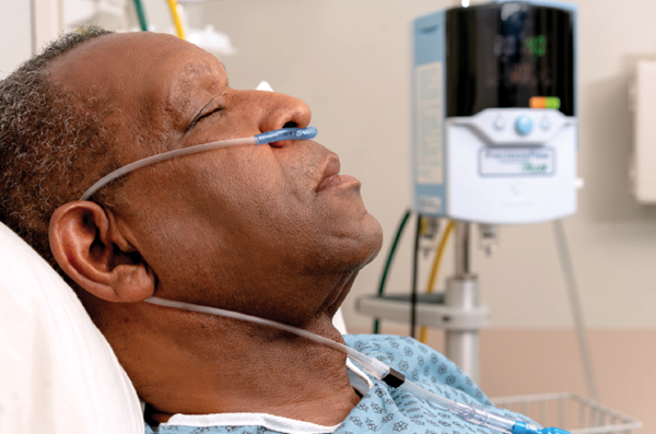
There are many perspectives available and early data to guide our management of COVID-19 patients that present with respiratory failure.
This article will review practice-altering data and the approach used by the authors at their institution with success. These hypoxic and crashing patients are difficult to manage, with the added challenge of provider safety being ever-present. The decision of oxygen delivery method, escalation of care, physiologic goals, and intubation procedure will be discussed below.
Background
COVID-19 is a viral respiratory infection caused by a positive sense RNA coronavirus that binds type 2 alveolar cells, intestinal epithelial cells, and vascular endothelial cells via the ACE2 receptor. This viral syndrome is marked by hypoxemic respiratory failure with decreased surfactant levels, direct cytopathic effect on pneumocytes, lymphocytic pneumonitis, and acute fibrinous organizing pneumonia leading to diffuse alveolar damage. Other aspects of this illness include a cytokine storm as well as disseminated intravascular coagulation with marked systemic inflammation and direct endothelial injury.8,11 The most likely and significant modes of transmission are droplet and contact transmission, with aerosol/airborne transmission being possible but less likely to contribute to severe disease burden.4 Infection prevention and control measures must include a variety of personal protective equipment, viral filters for respiratory support machines, consideration for aerosol-generating procedures (AGPs), and the possible role of drying agents or antimuscarinics. Goals in immediate care and resuscitation of the person-under-investigation (PUI) for COVID-19 in respiratory failure include decreased work of breathing, oxygen saturation greater than 90%,2 and improvement in mental status and other markers of end-organ perfusion.
Initial Evaluation and Measures
Patients should be transferred into a negative pressure room as soon as possible, receiving oxygen via a face-mask device if necessary (related to "dispersal distance" of potentially infectious droplets/aerosols specific to oxygen delivery methods reviewed below). Minimal personnel should be exposed to the patient at this point in care, including physician, nurse, and respiratory therapist, with other personnel donned with PPE available to help outside the room. The minimum PPE to be worn by health care workers in contact with the patient includes an inner and outer pair of gloves, gown, n95 or PAPR, and sealed goggles.
Choosing Oxygen Delivery Method
The key considerations in choosing an oxygen delivery method are the patient’s physiologic requirements and the risk of exposing providers to potentially infectious particles. The amount of oxygen deliverable (FiO2) and the dispersal distance of aerosols/respiratory droplets vary with each device and choices may also be institution-dependent. Based on high-fidelity mannequin studies, the following oxygen delivery methods are listed in order of increasing dispersal distance of aerosol; Non-rebreather mask (NRM), HFNC, nasal cannula, venturi mask, simple mask, nebulizer, NIPPV.3 The traditional nasal cannula can provide approximately 45% FiO2 with a dispersal distance of 40 cm. Venturi masks and simple masks provide approximately 50% FiO2 with dispersal distances of approximately 40 cm as well. NRMs provide FiO2 of approximately 90% and have a dispersal distance of only 10 cm, making this the ideal method of oxygen supplementation with regard to both amount of oxygen delivered and safety profile for healthcare workers. Many of these COVID-19 respiratory failure patients will require more than just additional oxygen, however. When supplemental O2 alone cannot improve the patient’s condition, one must consider high-flow oxygen systems (like the high flow nasal cannula [HFNC]) and non-invasive positive pressure ventilation (NIPPV) such as BiPAP or CPAP. HFNC can provide high FiO2 while boasting a dispersal distance of only 5-17 cm. This method decreases work of breathing and dead space while providing a small amount of positive end-expiratory pressure (PEEP). It involves oxygen-rich humidified gas with precisely set flow levels and oxygen concentrations.5 Importantly, the use of HFNC during the SARS-CoV outbreak in 2003 in Toronto did not contribute to the risk of HCW transmission.6 Even though HFNC seems to be the clear winner when choosing how to give extra oxygen, it must also be stressed that this device may still not be enough. Failure of HFNC may be indicated by the requirement of vasopressors, worsening RR and asynchrony, and possibly the ROX index greater than 4.88. The ROX index is a score created by Roca, et al. that combines respiratory rate, oxygenation status, and HFNC settings to generate an objective measure to predict failure of this form of non-invasive ventilation and indicate the need for endotracheal intubation. 7,1,10,9 NIPPV is not an ideal choice for the safety profile of HCWs. Dispersal distances up to 95cm or greater with BiPAP are intimidating, and if patients are requiring high levels of oxygen as well as positive pressure ventilation with clinical worsening, the patient may require endotracheal intubation to precisely control respiratory mechanics and reduce staff exposures. One way to minimize the viral dispersal is to use a BiPAP mask on a ventilator with viral filter in place on the expiratory limb to create a closed circuit, something that is not possible with BiPAP machines. Similarly, nebulized breathing treatments are also risky, and should be replaced with MDI treatments whenever possible.
Intubation
The use of a checklist method may be helpful in preparing for intubating patients, to minimize the opening of doors in negative pressure rooms as well as staff exposures.
- Preoxygenate: use a combination of HFNC with maximal settings with a NRB mask placed over the patient’s nose and mouth.
- Delayed sequence intubation vs Rapid sequence intubation: consider DSI if there is a concern for the ability to ventilate the patient once sedated and paralyzed. Ketamine should be the induction agent of choice for DSI and is a reasonable choice for RSI as well. Alternatively, etomidate can be used. Use rocuronium for paralysis for prolonged effect unless otherwise contraindicated.
- Fiberoptic intubation: avoid direct laryngoscopy when possible to reduce exposure during this AGP. An intubation box may be used over the patient as a physical barrier to respiratory droplets and aerosols. Consider leading with a bougie in anticipation of a difficult airway to increase the chance of first-pass success.
- Initial ventilator settings: LPV and APRV should be used to manage the significant atelectasis associated with COVID-19 respiratory failure and ARDS
Take-Home Points
The COVID-19 respiratory failure patient poses many challenging aspects of care.
- Healthcare workers are at risk of becoming infected when working in close proximity with patients, especially during AGPs.
- Liberal use of PPE and judicious use of personnel are key to reducing HCW exposures.
- The safest and most effective ways to provide respiratory support before endotracheal intubation are HFNC and NRB, which may be used in combination as well.
- Intubate with extreme caution using fiberoptic technology and consider DSI when appropriate.
References
- Bouadma L, Lescure F-X, Lucet J-C, Yazdanpanah Y, Timsit J-F. Severe SARS-CoV-2 infections: practical considerations and management strategy for intensivists. Intensive Care Medicine. 2020;46(4):579-582. doi:10.1007/s00134-020-05967-x
- Clinical management of severe acute respiratory infection ... https://www.who.int/docs/default-source/coronaviruse/clinical-management-of-novel-cov.pdf. Accessed July 20, 2020.
- Hui D.S., et al. Aerosol dispersion during various respiratory therapies: a risk assessment model of nosocomial infection to health care workers. Hong Kong Med J. 2014; 20(4):9-13. PMID: 25224111.
- Liu Y, Ning Z, Chen Y, et al. Aerodynamic analysis of SARS-CoV-2 in two Wuhan hospitals. Nature. 2020;582(7813):557-560. doi:10.1038/s41586-020-2271-3
- Nishimura M. High-Flow Nasal Cannula Oxygen Therapy in Adults: Physiological Benefits, Indication, Clinical Benefits, and Adverse Effects. Respiratory Care. 2016;61(4):529-541. doi:10.4187/respcare.04577
- Raboud J, Shigayeva A, Mcgeer A, et al. Risk Factors for SARS Transmission from Patients Requiring Intubation: A Multicentre Investigation in Toronto, Canada. PLoS ONE. 2010;5(5). doi:10.1371/journal.pone.0010717
- Rello, J., et al. High-flow nasal therapy in adults with severe acute respiratory infection: a cohort study in patients with 2009 influenza A/H1N1v. J Crit Care. 2012; 27(5):434-9. doi: 10.1016/j.jcrc.2012.04.006. Epub 2012 Jul 2.
- Ruan Q, Yang K, Wang W, Jiang L, Song J. Clinical predictors of mortality due to COVID-19 based on an analysis of data of 150 patients from Wuhan, China. Intensive Care Medicine. 2020;46(5):846-848. doi:10.1007/s00134-020-05991-x
- Roca O, Caralt B, Messika J, et al. An Index Combining Respiratory Rate and Oxygenation to Predict Outcome of Nasal High-Flow Therapy. American Journal of Respiratory and Critical Care Medicine. 2019;199(11):1368-1376. doi:10.1164/rccm.201803-0589oc
- Sztrymf B., et al. Beneficial effects of humidified high flow nasal oxygen in critical care patients: a prospective pilot study. Intensive Care Med. 2011. 37(11):1780-6. doi: 10.1007/s00134-011-2354-6. Epub 2011 Sep 27.
- Xu Z, Shi L, Wang Y, et al. Pathological findings of COVID-19 associated with acute respiratory distress syndrome. The Lancet Respiratory Medicine. 2020;8(4):420-422. doi:10.1016/s2213-2600(20)30076-x


