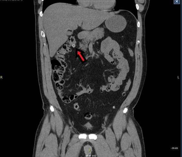
The epiploic appendix is a small pouch of peritoneum that protrudes from the serosal surface of the colon and is filled with fat and small vessels.
Acute epiploic appendagitis is an uncommon cause of abdominal pain and is due to torsion of the epiploic appendage or spontaneous venous thrombosis of a draining appendageal vein.1 This diagnosis can be clinically similar to sigmoid diverticulitis, and the diagnosis is often discovered after imaging on cross-sectional computed tomography (CT). CT imaging will show peripheral hyperattenuation with an area of fat stranding.2 Ultrasound findings demonstrate a fixed hyperechoic mass without Doppler color flow that is located adjacent to the anterior peritoneal wall and under the point of maximum pain.2
Case
A 45-year-old male presented to the emergency department with a chief concern of abdominal pain. He reported having 2 days of abdominal pain that began shortly after finishing lunch. Over the night prior to presentation, he experienced acute worsening of the pain and was unable to tolerate much oral intake secondary to pain. The pain was concentrated over the right flank and right lower quadrant pain and did not radiate into the groin. He denied any fevers, urinary symptoms, or vomiting. He had a history of nonalcoholic fatty liver disease and no previous abdominal surgeries.
Physical exam revealed an anxious-appearing male with normal vital signs. There was mild tenderness to palpation in the right lower quadrant. There was no rebound or guarding. He had normal male external genitalia with no overlying skin changes or tenderness to palpation of his scrotum. Cardiopulmonary examination was unremarkable.
Testing revealed a CBC and BMP within normal limits. AST and ALT were mildly elevated at 70 and 86, respectively. Lipase was within normal limits and a urinalysis was unremarkable. A CT with IV contrast of the abdomen and pelvis showed an ovoid area of mesenteric fat stranding and edema with central nodularity adjacent to the right hepatic flexure, most consistent with epiploic appendagitis. No focal fluid collection suggesting an abscess w visualized, though there were a few scattered colonic diverticula without evidence of acute diverticulitis. The patient was provided fluids, ketorolac, and ondansetron and was able to be discharged home with instructions for supportive care with pain management.
Discussion
More than 7% of patients presenting to the ED with symptoms clinically consistent with sigmoid diverticulitis are found to have primary epiploic appendagitis.1 Primary epiploic appendagitis is due to a torsion or venous thrombosis of the involved epiploic appendage.1 "Secondary epiploic appendagitis is associated with inflammation of adjacent organs, such as in cases of diverticulitis, appendicitis, or cholecystitis."3 Often acute epiploic appendagitis is misdiagnosed as diverticulitis or appendicitis, which can lead to unneeded hospitalization and invasive procedures. Epiploic appendagitis is associated with obesity, history of hernias, and exercise injuries. It is most commonly seen in males in their 40s.1,4
Clinical manifestations include acute abdominal pain, most commonly in the left lower quadrant. Patients are often afebrile and have unremarkable labs. CT imaging is the test of choice for diagnosis and findings include a "fat density ovoid lesion (hyperattenuating ring sign), mild bowel wall thickening, and a central high attenuation focus within the fatty lesion (central dot sign)."4
Management of acute epiploic appendagitis is conservative and symptoms often resolve in a few days.1,4 In a retrospective study of 660 CT scans performed for suspected diverticulitis or appendicitis, it was found that 11 scans (2%) showed features consistent with epiploic appendagitis, of which only 4 were correctly identified and 6 were misdiagnosed as diverticulitis and 1 was misdiagnosed as appendicitis.1 All of the misdiagnosed patients were hospitalized and 6 of the 7 received antibiotics.1 Though ED physicians rely on radiologists to identify findings suggestive of appendagitis, it is important to include this on the differential as misdiagnosis can result in inappropriate financial burden, misuse of antibiotics, and the possibility of an unnecessary surgery.4 Therefore, in cases where findings are indeterminate or questionably suggestive of an alternative diagnosis, it may be beneficial to discuss the possibility of appendagitis directly with the radiologist.
Take-Home Points
- >7% of patients with suspected sigmoid diverticulitis are diagnosed with epiploic appendagitis on CT.1
- Primary epiploic appendagitis is secondary to thrombosis or torsion of the epiploic appendix, while secondary is associated with inflammation of adjacent organs.3
- Management is conservative.
- Misdiagnosis may have significant unintended consequences.
References
- Rao PM, Rhea JT, Wittenberg J, Warshaw AL. Misdiagnosis of primary epiploic appendagitis. Am J Surg. 1998;176(1):81–5.
- Mollà E;Ripollés T;Martínez MJ;Morote V;Roselló-Sastre E;. Primary epiploic appendagitis: US and CT findings. Accessed December 01, 2020. https://pubmed.ncbi.nlm.nih.gov/9510579/
- Subramaniam, R. (2006, October). Acute appendagitis: Emergency presentation and computed tomographic appearances. Accessed December 01, 2020. https://www.ncbi.nlm.nih.gov/pmc/articles/PMC2579618/
- Suresh Kumar, V., Mani, K., Alwakkaa, H., & Shina, J. (2019, September 05). Epiploic Appendagitis: An Often Misdiagnosed Cause of Acute Abdomen. Accessed December 01, 2020. https://www.karger.com/Article/FullText/502683.


