THE CASE
The Patients
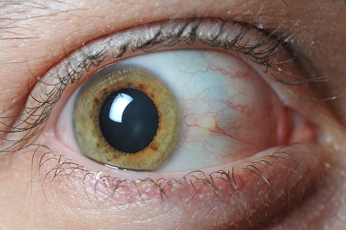
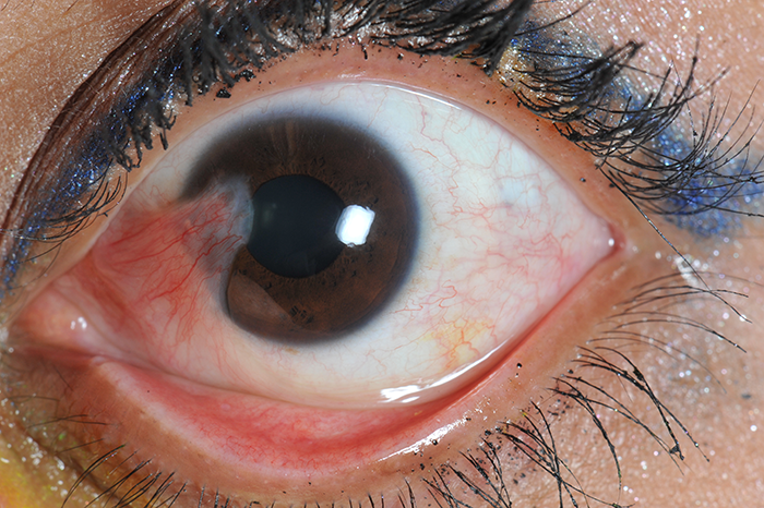
The images provided come from two different patients who presented to the ED with non-ocular complaints, but the ocular findings seen in the clinical photographs were noticed by the treating physician on physical examination. There is no history of direct eye trauma in either patient. Each patient reported an occasional foreign body sensation, which seemed to be more common in the summer months. Physical examination for both patients reveals visual acuity of 20/20 OD, 20/20 OS. Extraocular movements are normal. The ocular findings are chronic. What is your diagnosis for each patient?
Photo credits:
Patient A: Photo Courtesy of Rebecca Kasl, MSIV, Vanderbilt University School of Medicine
Patient B: Photo Courtesy of Tracey Hong, MSII, Vanderbilt University School of Medicine
What are the diagnoses?
THE DIAGNOSES
Patient A has a pinguecula, and Patient B has a pterygium.
A pinguecula is a lesion of the bulbar conjunctiva, and often appears as a partially translucent, colorless to light brown ridge adjacent to the limbus, usually on the nasal aspect. It may gradually enlarge with time, periodically become inflamed, or become a pterygium. A pterygium is a benign proliferation of fibrovascular tissue found in a triangular shape, extending across the limbus with the apex of the triangle pointing toward the center of the cornea.
Pterygia, though often asymptomatic, may also become inflamed, causing a foreign body sensation. A pterygium may cause decreased visual acuity if it encroaches on the visual axis, or if it exerts a mechanical deforming force on the cornea. Both disorders are more common in males and are associated with advanced age, exposure to ultraviolet light, and chronic eye irritation due to wind and dust.


