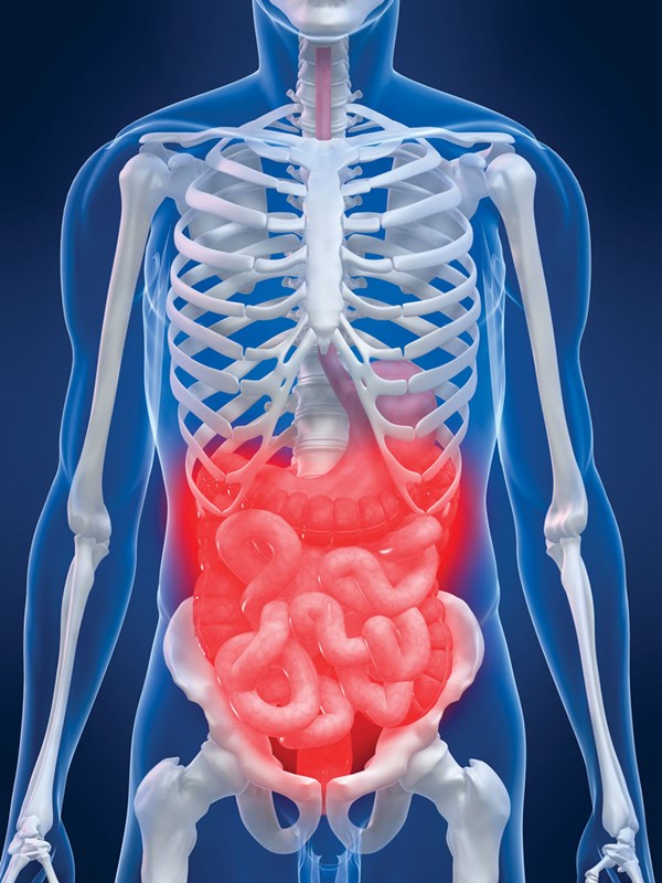
A 24-year-old male arrives to the emergency department (ED) after a motor vehicle collision. Initial workup reveals pelvic fractures with a small amount of retroperitoneal bleeding that is managed conservatively. A few hours later, the patient begins to feel very weak, develops oliguria, and complains of worsening abdominal pain. Physical exam reveals diffuse abdominal tenderness and distension. Lab work reveals worsening lactic acidosis and acute blood loss anemia. Intravesical pressures are found to be > 30 mmHg. The patient is diagnosed with abdominal compartment syndrome and is rushed to the operating room for abdominal decompression.
Abdominal Perfusion Pressure (APP) = Mean Arterial Pressure (MAP) — Intraabdominal Pressure (IAP)
Pathophysiology
Intraabdominal pressure (IAP) refers to the pressure within the abdominal cavity. The normal range is 5-7 mmHg, but obese and pregnant patients can have a normal IAP of up to 10-15 mmHg. Intraabdominal hypertension (IAH) is defined as a sustained IAP greater than or equal to 12 mmHg. Abdominal compartment syndrome (ACS) refers to a sustained IAP greater than 20 mmHg in the presence of end organ damage (Table 1).1 As IAP increases, abdominal perfusion pressure (APP) decreases, resulting in reduced blood flow and ischemia to visceral organs.
IAH can cause dysfunction of nearly every organ system:
Cardiovascular: IAH elevates the abdominal diaphragm, leading to a decrease in ventricular contractility and compliance. Obstruction of the inferior vena cava and associated increase in central venous pressure leads to a decrease in venous return to the heart. This results in decreased cardiac output (CO). It is not uncommon for patients to have persistent and significant sinus tachycardia in a last ditch effort to maintain CO.3
Pulmonary: Elevation of the abdominal diaphragm leads to atelectasis, decreased oxygen diffusion, increased intrapulmonary shunt fraction, increased alveolar dead space, and edema. In addition, patients with IAH that are also ventilated experience alveolar barotrauma due to increased peak inspiratory and mean airway pressure. This leads to further complications such as arterial hypoxemia, and hypercarbia.3
Renal: Decreased renal perfusion and acute renal failure is caused by renal vein compression and renal artery vasoconstriction, the latter induced by the sympathetic nervous and renin angiotensin systems in response to an overall decrease in blood flow to the kidney.3
Gastrointestinal: The effect of IAH on splanchnic organs leads to decreased gut perfusion, which can occur at pressures of just 20 mmHg. This causes intestinal ischemia and edema, further worsening IAP. This vicious cycle culminates in sepsis (due to loss of mucosal barrier and resultant bacterial translocation) and persistent multi-organ failure.4
Hepatic: IAH leads to a decline in the liver's ability to remove lactate. Increased lactic acid contributes to the metabolic acidosis often seen with ACS.4
CNS: IAH also has a direct effect on intracranial pressure (ICP). Sustained IAH can result in decreased cerebral perfusion and ischemia.5
Etiology
Risk factors for developing ACS can be divided into primary and secondary causes. Primary ACS is due to direct abdominal trauma, while secondary ACS is due to disease processes that do not necessarily originate in the abdomen (Table 2).1
Diagnosis
Because ACS often occurs in patients who are critically ill and unable to communicate, it becomes extremely important to understand and identify early warning signs of IAH.6
The three cardinal signs of ACS are worsening abdominal distension, difficulty breathing or elevated peak pressures on the ventilator, and decreased urine output. Other clinical signs include mental confusion, worsening hypoxia, hypotension, tachycardia, and jugular venous distention.
Measurement of the IAP is needed for definitive diagnosis, and the gold standard is measurement of bladder pressure using a Foley catheter and a transducer or manometer. Remember, ACS is not a diagnosis made on computed tomography (CT) scan! Several commercialized kits are available for the measurement of intravesical pressure, however an easy method that can be used in the emergency department involves instilling 50 ml saline into the bladder via the catheter, clamping the tubing of the collecting bag, inserting a needle through the specimen-collecting port and then attaching to a manometer.7
Treatment
The initial treatment for IAH includes addressing any underlying causes and initiating supportive treatment, which includes evacuation of intraluminal contents (rectal tube, nasogastric tube), evacuation of space-occupying fluid (eg, paracentesis for ascites), and measures to improve abdominal wall compliance, such as placing the patient in a reverse Trendelenberg position and ensuring adequate pain control and sedation.
Fluid resuscitation is extremely important for ACS patients as they often third space fluid and quickly drop their intravascular volume. This leads to hypotension which aggravates bowel ischemia and results in further third spacing and intravascular depletion.
For patients who do not improve with initial treatment, are unstable, or who have evidence of end organ damage, surgical decompression is the definitive treatment. Abdominal decompression is often performed in the operating room (OR) but also can be performed at the bedside if needed. Following decompression, patients are typically left open with a negative pressure wound therapy dressing. The patient usually returns to the OR in 1-3 days for a second look and possible fascial closure.8
Table 1. Key Differences between IAH and ACS.2
| Intraabdominal Hypertension (IAH) | Abdominal Compartment Syndrome (ACS) |
| IAP > 12 mmHg | IAP greater than 20 mmHg |
| No evidence of end organ damage | Evidence of end organ damage |
| Subclassified into grade 1-4 | Subclassified into primary and secondary causes |
Table 2. Primary and Secondary Causes of ACS
| Primary Risk Factors for ACS | Secondary Risk Factor for ACS |
| Pancreatitis | Trauma |
| Pelvic fractures | Burn patients |
| Liver transplant | Sepsis |
| Massive ascites | Septic shock |
| Bowel Distension | Hemorrhagic shock |
| Abdominal Surgery | Post-surgical patients requiring large volume resuscitation |
| Intraperitoneal bleeding | Ruptured abdominal aortic aneurysm, or pelvic fractures leading to retroperitoneal bleed |
Conclusion
As emergency medicine physicians, it is important to understand the signs and symptoms of life-threatening diagnoses such as ACS, which may be present or develop at any point along a high risk patient's ED course. Knowing the cardinal signs of ACS, using appropriate diagnostic tools to acquire IAP, and consulting surgery when ACS is suspected can save a patient's life.
References
- Malbrain ML, Cheatham ML, Kirkpatrick A, et al. Results from the international conference of experts on intra-abdominal hypertension and abdominal compartment syndrome. I. definitions. Intensive Care Med. 2006;32(11):1722-32.
- Shein M, Ivatury R. Intra-abdominal hypertension and the abdominal compartment syndrome. Br J Surg. 1998;85(8):1027-8.
- Cullen DJ, Coyle JP, Teplick R, Long MC. Cardiovascular, pulmonary, and renal effects of massively increased intra-abdominal pressure in critically ill patients. Crit Care Med. 1989;17(2):118-121.
- Quintel M, Pelosi P, Caironi P, et al. An increase of abdominal pressure increases pulmonary edema in oleic acid-induced lung injury. Am J Respir Crit Care Med. 2004; 169(4):534-541.
- Joseph DK, Dutton RP, Aarabi B, Scalea TM. Decompressive laparotomy to treat intractable intracranial hypertension after traumatic brain injury. J Trauma. 2004;57(4):687-93.
- Sugrue M, Bauman A, Jones F, et al. Clinical examination is an inaccurate predictor of intraabdominal pressure. World J Surg. 2002;26(12):1428-31.
- Melbrain ML. Different techniques to measure intra-abdominal pressure (IAP): time for a critical re-appraisal. Intensive Care Med. 2004;30(3):357-71.
- Kirkpatrick AW, Roberts DJ, De Waele J, et al. Intra-abdominal hypertension and the abdominal compartment syndrome: updated consensus definitions and clinical practice guidelines from the World Society of the Abdominal Compartment Syndrome. Intensive Care Med. 2013;39(7):1190-206.


