Case
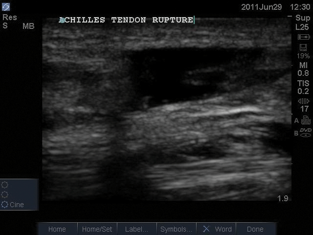
A 39-year-old male presents to the emergency department with pain and swelling in his right ankle two days after an injury he obtained while playing volleyball. He reports hearing a “pop” when landing after a jump, and has had swelling, difficulty walking and bearing weight, and difficulty with plantar flexion. His exam is remarkable for significant swelling, ecchymosis, and tenderness at the posterior aspect of the right ankle. He has weakness of right plantar flexion, but an equivocal Thompson test. He has no bony tenderness and is neurovascularly intact. Right ankle X-ray shows only evidence of swelling compatible with soft-tissue injury. Out of concern for a possible Achilles tendon rupture, a bedside ultrasound of the ankle is performed (Figure 1).
Failure to promptly diagnose an Achilles tendon rupture can result in retraction of the disrupted ends of the tendon, which may affect operative repair and result in prolonged disability.
Discussion
Achilles tendon rupture most often occurs after sudden dorsiflexion of a plantar-flexed foot, or by pushing off the foot with an extended knee. It is most commonly seen in the fourth or fifth decade of life, and may be related to repetitive overuse or deconditioned states.1 Traditionally, diagnosis has been made by history and physical examination. History often includes trauma with a “popping” sensation and sudden pain. Physical exam findings include the “hatchet strike” sign (a palpable proximal defect in the Achilles tendon) or a positive Thompson test.2 The area of rupture is likely to be found in the hypovascular area 3-6 cm proximal to calcaneal insertion.1
To perform the Thompson test, the examiner has the patient lie prone and squeezes the calf while observing the foot for slight plantar flexion. Pain and swelling can make diagnosis difficult. An estimated 20-25% are missed on initial exam, and are often misdiagnosed as ankle sprain.3 Failure to promptly diagnose an Achilles tendon rupture can result in retraction of the disrupted ends of the tendon, which may affect operative repair and result in prolonged disability.6 MRI is often used as a confirmatory test, but it is expensive, delays time to diagnosis, and there is no literature supporting its use.4
Point-of-care ultrasound (US) is becoming increasingly commonplace in the ED. With its low cost, expedience, and accessibility, US is finding expanding utility in musculoskeletal imaging. US offers a dynamic, physician-directed assessment of injuries that may otherwise only be seen with costly and time-consuming MRI exams. In the evaluation of suspected Achilles tendon rupture, US is a readily available and reliable means of diagnosis, and especially invaluable in patients with equivocal clinical exam findings.1,5
Some advocate US in the evaluation of suspected tendon injuries as the standard of care in the emergency department.6 Several clinical studies clearly demonstrate the effectiveness of US in the diagnosis of both partial and complete Achilles tendon injuries. US has a reported sensitivity of 96-100% and a specificity of 83-100%.1
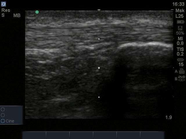
To perform the exam, the patient should be lying prone with the feet hanging off the bed. A high-frequency linear array probe is placed over the posterior ankle in long-axis, i.e., indicator pointed towards the patient's head. In long axis, the normal tendon has a fibrillar pattern with uniform thickness and echogenicity (Figure 2). In short-axis, the normal tendon has an oval shape with speckled echotexture. 7,8 The main US findings of Achilles tendon rupture are a loss of integrity of the path of the tendon, hematoma formation in the gap between torn tendon ends, posterior acoustic enhancement at the edges of the tear, and alterations in the echogenicity and shape of Kager's triangle (the pre-Achilles fat pad).1,7,9 Dynamic US can differentiate complete from partial ruptures. To perform this exam, have the patient gently plantar flex the ankle against resistance. A full-thickness rupture is indicated by movement of the tendon ends away from each other.1
Case conclusion
Bedside ultrasound clearly demonstrated a complete rupture of the Achilles tendon (Figure 1). The orthopedic service was consulted and a short leg posterior splint was placed. The patient was discharged with pain control, crutches, and instructions for non-weight bearing status. He was subsequently seen in the orthopedic clinic and scheduled for operative repair. In this case, bedside ultrasound enabled rapid evaluation and definitive diagnosis, thus expediting treatment and disposition in the face of somewhat equivocal physical exam findings.
References
- Adhikari S, Marx J, Crum T. Point-of-care ultrasound diagnosis of acute Achilles tendon rupture in the ED. Am J Emerg Med; 2012; 30(4): 634.e 3-4.
- Alfredson H, Masci L, Ohberg L. Partial mid-portion Achilles tendon ruptures: new sonographic findings helpful for diagnosis. Br J Sports Med; 2011; 45(5): 429-3.
- Daftary A, Adler RS. Sonographic evaluation and ultrasound-guided therapy of the Achilles tendon. Ultrasound Q. 2009; 25(3): 103-10.
- Kälebo P, Allenmark C, Peterson L, Swärd L. Diagnostic value of ultrasonography in partial ruptures of the Achilles tendon. Am J Sports Med. 1992; 20(4): 378-81.
- Maffulli N, Dymond NP, Capasso G. Ultrasonographic findings in subcutaneous rupture of Achilles tendon. J Sports Med Phys Fitness. 1989; 29(4): 365-8.
- Howard T, Butcher J. Achilles Tendon Rupture. The Little Black Book of Sports Medicine. 2006; 239-241.
- Ufberg J, Harris R, Cruz T, Perron, A. Orthopedic pitfalls in the ED: Achilles tendon rupture. 2004; 22(7): 596-600.
- Garras DN, Raikan SM, Bhat SB, Taweel N, Karanija H. MRI is unnecessary for diagnosing acute Achilles tendon ruptures: clinical diagnostic criteria. Clin Orthop Relat Res. 2012; 470(8): 2268-73.
- Bleakney RR, White LM. Imaging of the Achilles tendon. Foot Ankle Clin. 2005; 10(2): 239-54.


