


When trismus occurs, unconventional methods might be required to achieve and maintain the airway. This case highlights emergency medicine’s nimble approach to the difficult airway.
Case Report
During a night shift, the ED team is called to the floor for emergent intubation of an admitted patient due to sudden hypotension, altered mental status, and inability to protect his airway. The patient is a 56-year-old male with a history of rheumatoid arthritis on prednisone and methotrexate, admitted 2 weeks ago for management of enterococcus faecalis bacteremia, COVID pneumonia, and influenza. Pre-intubation evaluation of the patient reveals an obese, edentulous man able to open his mouth greater than 3 finger widths.
Procedure
Blood pressure demonstrated a MAP of 50 and therefore the patient was not immediately intubated but rather first resuscitated with norepinephrine. He was able to be oxygenated with bag valve mask ventilation and jaw-thrust maneuver throughout this process and was transferred from the floor to the ICU. After his blood pressure normalized, he was given etomidate and rocuronium for rapid sequence intubation. After waiting approximately 1 minute for the rocuronium to take effect, the patient became apneic but developed trismus and his mouth was unable to be opened, deviating from his previous airway exam.
With concern for IV infiltration causing failure of medication delivery, another dose of rocuronium was administered, this time via a separate IV site. Once again, despite waiting a minute, the patient demonstrated trismus, with a maximum jaw opening of approximately 20 mm. Palpation of the temporomandibular joint (TMJ) did not demonstrate any abnormalities in the mandibular fossa, although this assessment was challenging because of the patient’s body habitus. Despite these challenges, ventilation and oxygenation were able to be maintained via BVM; therefore, cricothyrotomy was not attempted.
Fiberoptic video bronchoscopy was then attempted. The bronchoscope was inserted through the lateral aspect of the mouth and revealed an edematous airway obscuring the view of the vocal cords. The patient’s tongue was sutured to the side to allow insertion of a video laryngoscope, which again revealed a partial view of an edematous supraglottic airway. An ET tube could not be passed through the narrow jaw opening, and the video laryngoscope was removed. An LMA was then able to be inserted through the small opening. The connector cap was removed, and a video bronchoscope was inserted through the LMA with subsequent visualization of an edematous glottis partially obstructing the vocal cords. The disposable bronchoscope was then cut and used as an introducer to pass a size 6.0 ETT through the cords. Video laryngoscope was removed and both LMA and ETT were left in place. Follow-up chest x-ray demonstrated appropriate position of the ETT within the LMA.
Five days later, the ICU team was able to remove the LMA (but kept the ETT in place). The patient’s trismus continued throughout the rest of his hospitalization, and he was not extubated. CT maxillofacial bones did not demonstrate any abnormalities of the TMJs or mandible. The oral maxillofacial surgery team was consulted with concern for trismus; however, prior to their evaluation, the patient unfortunately passed away due to worsening septic shock.
Figure 2. Video laryngoscope view of the supraglottic cavity with vocal cords obscured by swelling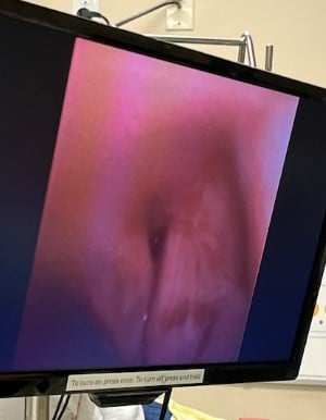
Background
"Trismus" is defined as limited jaw opening less than 35-55 mm. In a normally functioning jaw, the muscles of mastication (the medial pterygoid, lateral pterygoid, temporalis, and masseter muscles) attach to the rami of the mandible and contract to close the jaw via innervation by CN V3. Specifically, these muscles coordinate the hinge movement of the condyle in the mandibular fossa as the jaw begins to open. As the jaw opens further, the condyle glides anteriorly, resulting in a fully open mouth. Between the fossa and the condyle, an articular disc serves as cartilage. A problem with any one of these mechanisms may result in trismus.1
Figure 3. In normal jaw opening, the mandibular condyle sits in the mandibular fossa and initially rotates and then translates anteriorly over the anterior disc (blue) 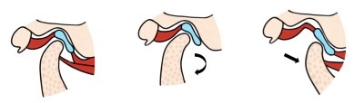
Figure 4. In trismus, the anterior disc can be deranged and the mandibular condyle cannot rotate or translate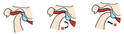
The presence of trismus is assessed prior to intubation by placing 3 fingers in the patient’s mouth. Trismus is considered to be present if the jaw cannot open wider than 3 finger widths. The evaluation of trismus is a component of the 3-3-2 rule, which serves as a quick method to predict a difficult airway. Apart from mouth opening, hyoid-mental distance is measured as 3 fingerbreadths, and thyroid mental distance is measured as 2 fingerbreadths. Any deviation in these brief measurements constitutes a difficult airway that may require cricothyrotomy, nasal intubation, or other advanced techniques that may be more safely done in the operating room.2
Discussion
In this report, we discuss the hazard of developing trismus peri-intubation and unconventional methods used to intubate. Past case studies report on trismus before intubation or following extubation, but review of the literature reveals very few cases of trismus developing during intubation.
We considered many different etiologies of acute trismus. Rocuronium (of which we gave a total of 200 mcg) should have provided an adequate neuromuscular blockade to combat any sort of mechanism of trismus that occurred secondary to events at the motor end plates. These include upper motor neuron lesions (patient did have an additional history of strokes), and seizures. Hypocalcemic tetany was considered; however, ABG demonstrated a normal ionized calcium. Etomidate was considered, as side effects include spasm; however, the effects are usually transient and do not last for 5 days.
We also considered the possibility that succinylcholine was given in the place of rocuronium erroneously. This drug can cause malignant hyperthermia with associated muscle rigidity that can be refractory to neuromuscular blockade, but the absence of subsequent fever makes this diagnosis unlikely. A side effect of succinylcholine is also muscular spasms, but once again, we would have expected this side effect to have resolved several minutes following administration. There have been case reports of nondepolarizing agents, such as rocuronium, causing muscular rigidity, but these are very rare, and are thought to occur in patients predisposed to malignant hyperthermia via ryanodine receptor mutations.3 Both drugs are present in the Emergent Intubation Medication box taken to floor intubation; however, inspection and medication counts confirmed rocuronium had been administered.
We considered extensive supraglottic swelling, believed to be as a result of COVID and influenza infection in this patient, could have contributed to the trismus, as trismus is a known chief complaint of supraglottic swelling.4 An allergy to one of the intubation medications contributing to pre-existing soft tissue swelling was also considered; however, this is unlikely as patient did not display other physical exam findings of an allergic reaction.
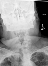
Figure 5. Narrowed airway seen on x-ray
It is possible that the jaw-thrust maneuver, which was held for around 30 minutes in order to maintain oxygenation as the patient was resuscitated with norepinephrine and transferred to the ICU, caused a TMJ abnormality that resulted in trismus. Displacement of the anterior disc is a known mechanism of lock jaw, as are mandibular fractures. Pre-existing joint space disease caused by the patient's rheumatoid arthritis (the TMJ is affected in 22% of those with rheumatoid arthritis) may have predisposed the patient to sustaining excessive trauma via the jaw-thrust maneuver such that either one or both of the anterior discs were displaced.5 A mechanical abnormality of the jaw is likely given the trismus did not resolve with neuromuscular blockade or the passage of time. CT maxillofacial was completed following intubation, and the radiologist did not comment on any TMJ bony abnormalities; however, MRI is the gold standard to evaluate the anterior discs as such small cartilaginous abnormalities may be missed on CT.5
Conclusion
This case highlights that trismus can occur during intubation and that unconventional techniques (eg, an ETT through a laryngoscope) may be required to allow for a successful intubation. Emergency physicians should be aware that pre-existing rheumatoid disease may affect the TMJ and prepare for the possibility of a difficult airway in these patients.
References
- Rodhri Davies LC. Case 8: Painful trismus. In: Avijit Banerjee ST, ed. Odell’s Clinical Problem Solving in Dentistry. Vol 4. Elsevier; 20220:37-40.
- al RWe. Airway. In: Rosen’s Emergency Medicine: Concepts and Clinical Practice E-Book. Vol 10. Elsevier; 2022:2462.
- Albrecht A, Wedel DJ, Gronert GA. Masseter muscle rigidity and nondepolarizing neuromuscular blocking agents. Mayo Clin Proc. 1997;72(4):329-332.
- Aiello G, Metcalf I. Anaesthetic implications of temporomandibular joint disease. Can J Anaesth. 1992;39(6):610-616.
- O'Connor RC, Fawthrop F, Salha R, Sidebottom AJ. Management of the temporomandibular joint in inflammatory arthritis: Involvement of surgical procedures. Eur J Rheumatol. 2017;4(2):151-156.