


THE CASES
Case 1. The Patient
A 15-year-old female presents to the ED for the fourth time in three weeks because of low-grade fever, malaise, and chest pain. Over the past day she developed a painful skin eruption on the right index fingertip pad. The patient has a mild headache and diminished visual acuity in the right eye. Physical examination reveals an ill-appearing patient with a temperature of 101 °F and heart rate of 110 beats per minute. Funduscopic examination of the right eye shows a white, centered retinal hemorrhage lateral to the disc. A II/VI systolic murmur is heard.
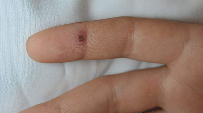
What is the lesion in the image and what is the diagnosis?
Case 2. The Patient
A 41-year-old male presents to the ED for three days of fever and painless skin eruption to the left palm. The patient has been known to inject feces into his veins, and has lost fingertips due to this behavior. Physical examination reveals a mildly ill patient with a temperature of 100.6 ° F and heart rate of 117 beats per minute. A II/VI systolic murmur is heard.
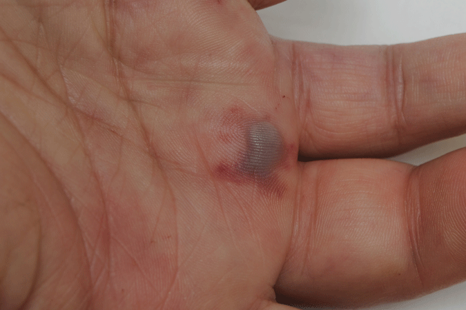
What is the lesion in the image and what is this diagnosis?
The Diagnosis
Case 1. Osler's Node/Infective Endocarditis
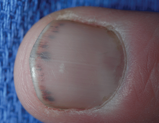
This patient has acute infective endocarditis (IE). The fingertip eruption is an Osler's node, named after Sir William Osler. The finding is due to an embolic manifestation of IE that initially is a micro-abscess that becomes sterile and propagates an immune-mediated vasculitis giving these lesions a palpable purpura or nodular feature. Osler's nodes are purpuric, slightly raised and tender, and are typically found on the pads of the fingers or toes. They may occur at any time during the course of IE, and last from hours to days. Splinter hemorrhages though less specific for IE, are thin, brownish-red eruptions underneath the fingernails that follow the direction of nail growth. They represent immune complex deposition in small peripheral vessels causing a vasculitis.
Case 2. Janeway Lesion/Infective Endocarditis
Patient 2 also has IE. The palmar eruption is a Janeway lesion which is an embolic manifestation of IE frequently caused by Staphylococcal aureus, and histologically appears as subcutaneous abscess. Bacteria can be cultured from the eruption (which is a painless), irregular erythematous macule, or patch on the palms or soles. Janeway lesions may persist for days to weeks.
Photos courtesy of Lawrence B. Stack, MD and R. Jason Thurman, MD