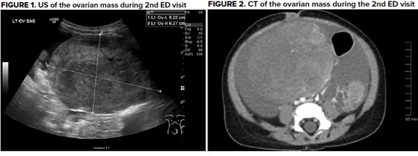
This case study describes the presentation of an 8-month-old female who had been misdiagnosed with constipation twice in the previous 36 hours before being correctly diagnosed with ovarian torsion and a tumor, identified as a Juvenile Granulosa Cell tumor.
Author's note: Author is father of the patient. No other disclaimers.
The overall incidence of granulosa cell tumors varies from 0.4 to 1.7 cases per 100,000 women.1 The juvenile form of this tumor is even rarer, being only 5% of these cases. The incidence of ovarian torsion among females ages 1-20 years is estimated to be 4.9 of 100,000.2
The purpose of this case report is to show the importance of using appropriate imaging before settling on a diagnosis.
Case Presentation
An 8-month-old female with no past medical history presents to the pediatrician with abdominal distention (noticed by parents as having tighter clothes/diaper and visually large abdomen), fever up to 103°F, decreased urinary output, and decreased activity. This physician diagnosed the patient with constipation after ordering an abdominal x-ray to look for stool burden. This was read as normal. A few hours later, the symptoms worsened, so the patient was brought to a pediatric ED. No imaging was obtained. The patient was again diagnosed with constipation and discharged. The following day, the patient developed increased work of breathing, livedo reticularis, and increased abdominal distention. There had been no urinary output for 24 hours. She was brought to a different pediatrician who observed lack of bowel sounds. The patient was then referred to a different pediatric ED than the day before.
At the ED, triage showed a temperature of 39.9°C and RR 44. Initial labs showed a WBC of 23.8, hemoglobin of 9.4, platelet of 720, and CRP of 190. Labs and vitals were otherwise unremarkable. Abdominal exam was documented as "soft, no organomegaly, abdominal distention, bowel sounds absent. Moderated diffuse tenderness throughout." A formal ultrasound was ordered to rule out intussusception. This revealed a mass measuring 9.2 x 7.5 x 6.3cm in the lower abdomen, likely arising from the left ovary, as well as a large amount of ascites (seen in Figure 1). This was confirmed with CT (seen in Figure 2). The CT also suggested ovarian torsion.
Management and Outcome
The patient was brought to the operating room for diagnostic laparoscopy and ovarian detorsion. 400cc of bloody ascites were also drained. Three days later, an additional 800cc of bloody ascites were drained, and removal of the mass and ovary was performed. The mass was identified as a juvenile granulosa cell tumor, and the patient was referred to the local pediatric oncological hospital.
Discussion
This case demonstrates the importance of point of care ultrasound in the ED. Because no imaging was obtained during the first ED visit, the patient had to experience an additional 24 hours of discomfort caused by ovarian torsion secondary to the mass. The delayed diagnosis could also have contributed to a prolonged hospital course and increased time in the PICU. Also, an x-ray might not be sufficient imaging to implement in a case of abdominal distention, as it proved to be ineffective in this case. Abdominal x-ray has sensitivity/specificity of 82/83% for small bowel obstruction3 and 84/72% for constipation.4
It is a well-established practice to use abdominal x-ray as an initial diagnostic test, but if this proves to be insufficient in providing a diagnosis, an ultrasound is a reasonable next step in workup. Ultimately, POCUS is fast and cheap and should be integrated into pediatric emergency medicine training just as it has in general EM training. Using POCUS in cases of abdominal distention could help prevent misdiagnosis.
References
- Calcaterra V, Nakib G, Pelizzo G, et al. Central precocious puberty and granulosa cell ovarian tumor in an 8-year old female. Pediatr Rep. 2013;5(3):e13.
- Guthrie BD, Adler MD, Powell EC. Incidence and trends of pediatric ovarian torsion hospitalizations in the United States, 2000–2006. Pediatrics. 2010;125:532-538.
- Thompson WM, Kilani RK, Smith BB et al. Accuracy of abdominal radiography in acute small-bowel obstruction: does reviewer experience matter? AJR Am J Roentgenol. 2007;188(3):W233–W238.
- Jaffe T, Thompson W. Large bowel obstruction in the adult: Classic radiographic and CT findings, etiology, and mimics. Radiology. 2015;275(3):651-663.


