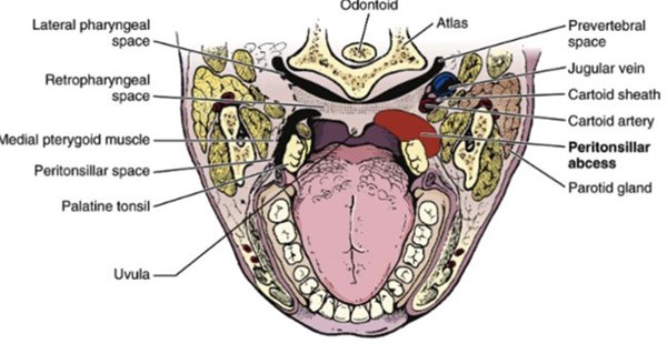
A peritonsillar abscess (PTA) is a deep oropharyngeal infection that results in an accumulation of purulent material in the potential space between the tonsillar capsule and the superior constrictor muscle.1 This is often secondary to tonsillitis that progresses to tonsillar capsule rupture and peritonsillar cellulitis. PTAs are most commonly seen in adolescents and young adults with 80% of cases occurring between ages 10-40.2,3 Clinical exam findings for PTA include sore throat with unilateral swelling and uvular deviation, “hot potato voice,” fever, trismus and inability to tolerate secretions.1-3 If untreated these infections may lead to extension into the mediastinum, erosion into vasculature (Lemierre’s syndrome), or spontaneous rupture into the pharynx causing aspiration.4 In order to avoid these complications prompt diagnosis, drainage, and antimicrobial treatment must be provided.
Unfortunately, history and physical exam alone are unreliable when diagnosing a PTA. Even when performed by otolaryngologists (ENTs), physical exam has been shown to only have a sensitivity of 78% and specificity of 50%.2 Blind needle aspiration is also a method of diagnosis but is unreliable as well with a false-negative rate of 10% to 24%.3 Computerized tomography (CT) studies with contrast on the other hand have a sensitivity of 100% but are not without their risks.3 With the highest rates of incidence in young populations, radiation exposure makes increasing cancer risk a real threat.
Because of this, ultrasound (US) has gained traction in the diagnosis and treatment of PTAs. Recent studies have indicated that ultrasound imaging is a safe and effective alternative to CT scans.2,5 Currently two methods exist to evaluate for a PTA: intraoral and transcutaneous. The intraoral approach has been more commonly used and has demonstrated a slightly higher sensitivity and specificity and will therefore be the focus of our discussion.
Intraoral US has a sensitivity ranging from 89-95.2% and specificity of 78.5-100%, which approaches that of contrasted CT scans.2 Intraoral US has been shown to decrease overall cost of care and one study demonstrated a decreased ED length of stay by 66 minutes.2,6 The use of US not only allows the ED provider to rapidly diagnose a PTA but also enables direct visualization of the needle during the procedure to ensure drainage. When compared to the landmark aspiration approach performed by ED providers, the use of US decreased the need for ENT consultation from 50% to 7% and had higher successful drainage rates.6 Complications of blind and US guided needle drainage include aspiration, hemorrhage, and puncture of the internal carotid.1
Technique
Prior to ultrasounding the suspicious area it is essential to obtain local anesthesia, especially in patients with a strong gag reflex. This may be performed through topical gel lidocaine, nebulized lidocaine, benzocaine spray, or injected anesthetics. We recommend topical benzocaine spray due to ease of provider use and patient comfort. To avoid aspiration, have the patient sitting up in the bed with suction and a spit basin available. To improve visualization of the posterior pharynx the provider may utilize a tongue depressor or ideally a Macintosh blade inserted to the base of the tongue while the patient holds the handle for improved control and light access.
The probe of choice for intraoral PTA drainage is currently the endocavitary probe. The probe should be obtained from sterile processing and a protective cover should be placed over the probe, which may be a condom, sterile probe cover or the finger of a standard glove. Prior to insertion of the probe into the cover ensure that gel is applied inside. It is not necessary to have gel on the outer surface due to the moist mucous membrane being targeted. If required sterile gel is preferred, however. If marked trismus is present using a smaller linear array known as the “hockey stick” intraorally or attempted visualization through the transcutaneous approach under the mandibular angle with a linear probe may be performed.7
Insert the probe in the transverse orientation starting from the contralateral side of the mouth to the ipsilateral tonsil. Begin with visualization of the unaffected side to determine normal anatomy prior to inspection of the affected side. Be aware that PTAs may occur bilaterally in <10% of cases.1 The abscess can be identified as a hypoechoic collection between the tonsil and lateral oropharynx. It is essential to fan superior to inferior as 70% of PTAs occur in the superior pole and 30% in the inferior pole.3 When the PTA is identified, measure it in two planes to estimate volume. Next determine the depth from the surface, average being 9 mm. Once identified locate the internal carotid artery. The internal carotid location ranges 5-25 mm posterior to the abscess wall. Color flow may aid in visualizing the vasculature.3
The plastic needle guard from an 18-gauge needle may be cut to the depth of the abscess and reinserted over the needle to help prevent damage to deep vasculature. For smaller abscesses, a 3 mL syringe is ideal to decrease resistance while improving dexterity in the oral cavity. For larger abscesses a 5 or 10 ml syringe may be necessary. The needle should be inserted lateral to the probe and visualized using an in-plane approach to keep the needle tip in view through the entire procedure. Constant negative pressure should be applied to the syringe. The provider should be able to visualize the abscess being drained. Once obtained, the purulent material should be sent for cultures. After drainage the patient’s clinical picture should be taken into consideration when determining the need for IV antibiotics as an inpatient or if stable for discharge on oral antibiotics and close ENT follow-up.
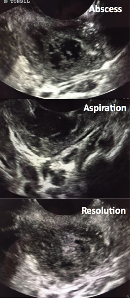
FIGURE 1. Ultrasound Visualization
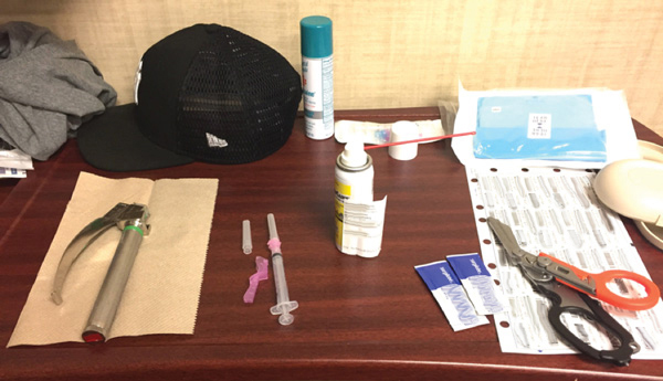
FIGURE 2. Setup
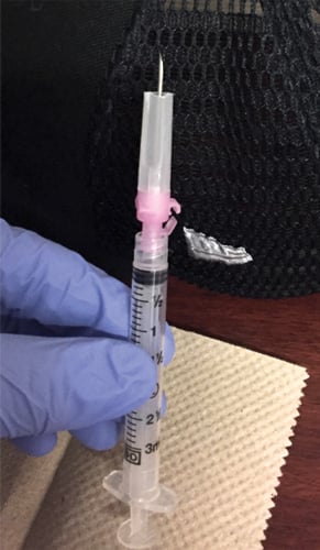
FIGURE 3. Needle Preparation
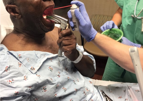
FIGURE 4. Patient Positioning and Prep Limitations
Pediatric populations may be unable to tolerate intraoral bedside ultrasound and drainage, thus requiring either transcutaneous US or a CT scan. Operative drainage may be necessary. The learning curve for US diagnosis of PTAs at bedside has been shown to be short requiring 3 to 4 patient experiences.3 If uncomfortable with the procedure, an experienced provider or an ENT specialist should be present at bedside. If there is concern for extension of the infection or unable to confidently visualize the PTA on US, a CT scan should be performed for further characterization.
Conclusion
The use of bedside US has continued to expand the diagnostic and procedural capabilities of the EM provider. Intraoral US has been found to be a safe and cost effective way to diagnose and treat PTAs in the ED. It has demonstrated the ability to decrease patient length of stay in the ED and to drastically reduce the need for specialist consultation. With the continued rise in ED patient visits, emphasis on throughput, and focus on medical cost saving this should be a procedure in every EM provider’s skill set.
References
- Mapalli E, Sabhaney V. Stridor and Drooling in Infants and Children. In: Tintinalli’s Emergency Medicine: A Comprehensive Study Guide. 8th ed. New York, N.Y.: McGraw-Hill Education LLC; 2016:800-801.
- Froehlich MH.; Huang Z; Reilly BK. Utilization of ultrasound for diagnostic evaluation and management of peritonsillar abscesses. Curr Opin Otolaryngol Head Neck Surg. 2017;25(2):163-168.
- Secko M, Sivitz A. Think ultrasound first for peritonsillar swelling. Am J Emerg Med. 2015;33(4):569-572.
- Brook I. Microbiology and management of peritonsillar, retropharyngeal, and parapharyngeal abscesses. J Oral Maxillofac Surg. 2004;62(12):1545-50.
- Lyon M, Blaivas M. Intraoral ultrasound in the diagnosis and treatment of suspected peritonsillar abscess in the emergency department. Acad Emerg Med. 2005;12(1):85-88.
- Costantino TG, Satz WA, Dehnkamp W, Goett H. Randomized trial comparing intraoral ultrasound to landmark-based needle aspiration in patients with suspected peritonsillar abscess. Acad Emerg Med. 2012;19(6):626-631.
- Prokofieva A, Modayil V, Chiricolo G, Ash A, Raio C. Ultrasound-guided drainage of peritonsillar abscess: shoot with your hockey stick. Intern Emerg Med. 2016;11(6):883–884.


