We present a case of a 41-year-old male with a history of IV drug use and MRSA bacteremia who presented to the ED with a rash and bilateral vision loss.
The patient endorsed a rash in the upper and lower extremities, which worsened over the last several months. In addition, he endorsed several weeks of a non-painful rash on his scalp with associated hair loss. Regarding his visual complaints, his symptoms began several weeks ago as night vision impairment. Over time, his vision continuously worsened, and upon presentation, he had developed significant "cloudiness" at the center of his visual fields. He reported only seeing “outlines” of objects or people, but his vision overall was very hazy. A brief review of systems was significant for bilateral ocular discharge, headache, as well as a genital sore which resolved several weeks ago. No fevers were reported.
Past medical history included acute septic pulmonary embolism, allergic rhinitis, anxiety, and GERD. He reported using albuterol and eucerin, and listed allergies to cephalosporins, penicillin, and pollen.
The patient endorsed a long history of IV drug use (methamphetamine), most recently within the past month. The patient also states his last sexual encounter was over a year ago. He was unemployed and living with his father. He denied ethanol use but reported smoking 2-4 cigarettes monthly. He denied recent travel.
Upon physical exam, the patient appeared well-developed, pleasant, in no apparent distress, with no focal deficits neurologically. Vital signs were BP 138/84, pulse 109, RR 20, temp 99.7, oxygen 97 % on room air.
The patient’s eyes showed traces of edema on both lids/lashes, with chemosis/injection with moderate clear/yellow discharge. His corneas were mildly hazy, without defects or edema. Anterior chamber was 4+ flare, cells suspended; irises showed synechiae with prominent vessels. The patient’s lenses showed pigment on the capsule, with hazy anterior vitreous. Extraocular movements were intact. Acuity in the right was sensitive to hand motion, and in the left, to light perception.
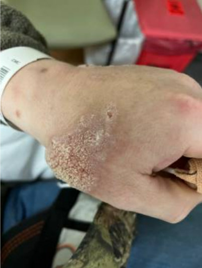
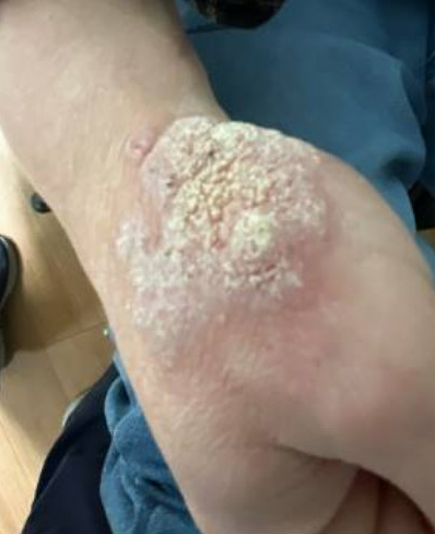
The patient’s skin showed multiple irregular rough, scaly plaques over bilateral upper extremities, and irregular erythematous macules over the anterior scalp with overlying fine scale. Bilateral lower extremities presented with multiple irregular erythematous macules with areas of excoriation.
The differential diagnosis included lupus, syphilis, HIV, HPV, gonorrhea/chlamydia, HLA-B27 related condition, hypertensive retinopathy, diabetic retinopathy, bacterial/viral conjunctivitis, uveitis, endophthalmitis, verruca dermatitis, cutaneous syphilis, and other etiologies.


Initial Evaluation
Initial workup from the emergency department revealed elevated CRP (12), ESR (109), and an unremarkable CBC and BMP. Multiple laboratory tests were subsequently added on including blood cultures, ANA , quantiferon, Toxoplasma Gondii IgG/IgM Antibody, and Histoplasma, all of which were negative. Given the severity of the vision loss and the extensive rash, consultations were requested from Dermatology and Ophthalmology.
The patient was evaluated by Dermatology, who believed the scalp rash was likely consistent with seborrheic dermatitis, but that the bilateral upper and lower extremity rash was felt to be consistent with very large verrucae. Given the unusual size and persistence of verrucae, they suspected the patient was likely immunosuppressed, and additional labs were ordered including HIV (HIV-1 positive, RPR (reactive, >1:1024), FTA ABS (reactive), eye cultures (negative).
The patient was also seen initially in the ED by Ophthalmology, who then brought the patient to their clinic for a more detailed eye examination outlined above. Their findings were concerning for endophthalmitis, and the patient was returned to the ED for admission to the Hospitalist service, with Dermatology, Ophthalmology, and Infectious Disease consulting.
Confirmatory Evaluation/Diagnosis
At the Ophthalmology clinic, where their examination was concerning for bilateral endogenous endophthalmitis, an ocular paracentesis was performed and the patient was instructed to return to the ED for admission for IV antibiotics, ID consultation, and an echocardiogram to rule out endocarditis. During his admission, labs were significant for reactive RPR/FTA-ABS, HIV-1 antibody, and CMV IgG. Blood cultures showed no growth and fungal culture was negative. Hepatitis B, Hepatitis C, ANA, and ANCA were negative. His toxicology screen was positive for methamphetamine. CSF following a lumbar puncture demonstrated elevated protein and reactive VDRL. Echocardiogram was negative for endocarditis. Eye culture demonstrated rare PMNs. A left forearm skin biopsy was consistent with secondary syphilis.
Diagnosis and Outcome
Based on the physical exam and laboratory findings, the patient was diagnosed with syphilis meningitis, ocular syphilis (endophthalmitis), and syphilitic dermatitis, in the setting of new HIV infection. The patient was admitted to the hospital for 2 weeks of IV penicillin. He received a dose of intravitreal ceftazidime as well as voriconazole as well. Around the time of discharge, the patient was endorsing a persistent but improved visual deficit. Repeat visual acuity was significant for the following:
- Visual Acuity
- Right: 20/200
- Left: HM/inconsistent CF
- Adnexa/Lids
- Right: Mild crusting
- Left: Mild crusting
- Conjunctiva/Sclera
- Right: White and quiet
- Left: trace injection
- Cornea
- Right: clear
- Left: clear
- Anterior Chamber
- Right: Grossly quiet
- Left: improving clot of fibrin/debris in front of the lens, tr hyphemia in area of clot
- Iris
- Right: Dilated, all synechiae broken
- Left: Irregular, breaking synechiae but still attached to fibrin clot, yellowed iris
The patient was subsequently discharged with ophthalmology and addiction medicine referrals. He was provided prescriptions for steroid eye drops and started on bictegravir/emtricitabine/tenofovir alafenamide.
Management
Syphilis is known to progress through 4 different stages, each with unique signs and symptoms. During primary, secondary, or early latent syphilis the patient can often be cured with a single injection of long-acting benzathine penicillin-G. Late latent syphilis is treated with 2.4 million units of penicillin G once a week for three weeks. Once a patient develops syphilitic meningitis or ophthalmic syphilis, the treatment includes IV penicillin for 10-14 days.1
The hallmark of HIV treatment involves antiretroviral therapy. There are several classes of antiretroviral medications and regimens including nucleoside reverse transcriptase inhibitors, integrase strand transfer inhibitor-based regimens, non-nucleoside reverse transcriptase-based regimens, and protease inhibitor-based regimens. While these medications cannot cure HIV or AIDS they are successful in attaining treatment goals which include maximally and durably suppressing plasma HIV RNA, the restoration and preservation of immunologic function, and the reduction of HIV-associated morbidity. This therapy also serves to prolong the duration and quality of survival, and finally to prevent HIV transmission.
Discussion
Human immunodeficiency virus is generally acquired through sexual intercourse but may also be passed perinatally or through exposure to infected blood (IVDU, health care-related exposure). Acute HIV may be associated with vague symptoms such as fever, rash, diarrhea, and headache; however, 60% of individuals will be asymptomatic. The HIV attaches to the CD4 molecule and CCR5, which allows the virus to fuse and spill contents into T-cells. The virus integrates into the host genome, which eventually leads to viral budding and the release of more viruses.
Over time, a patient’s T-cell count will drop, leaving them susceptible to successive AIDS-defining illnesses. The hallmark of treatment for HIV is known as highly active antiretroviral therapy (HAART). HAART often consists of drugs from at least two different classes of antiretroviral therapy such as nucleoside analog reverse transcriptase inhibitors, non-nucleoside analog reverse transcriptase inhibitors, and protease inhibitors.2 Current recommendations call for HAART as quickly as possible after the diagnosis is made. For those who are diagnosed in the setting of an opportunistic infection, HAART should be initiated shortly after the infection has been adequately treated.2
Syphilis has been found to enhance the transmission of HIV, likely secondary to increased incidence of genital ulcerations, which promote spread. HIV patients are more likely to present initially with secondary syphilis as well as more rapid progression to neurosyphilis.3 An LP is required for the diagnosis and patients found to be positive require an LP every 6 months until negative.4 Early treatment of these patients is imperative, and the treatment of neurosyphilis requires nearly 2 weeks of IV penicillin.
References
- French P. Syphilis. BMJ (Clinical research ed.) 2007;334(7585):143–147.
- Gandhi RT, Bedimo R, Hoy JF, et al. Antiretroviral Drugs for Treatment and Prevention of HIV Infection in Adults: 2022 Recommendations of the International Antiviral Society–USA Panel. JAMA. 2023;329(1):63–84.
- Ha T, Tadi P, Dubensky L. Neurosyphilis. [Updated 2023 Jul 3]. In: StatPearls [Internet]. Treasure Island (FL): StatPearls Publishing; 2024.
- Lewis DA, Young H. Syphilis. Sex Transm Infect. 2006 Dec;82 Suppl 4(Suppl 4):iv13-5.


