Compartment syndrome is an orthopedic emergency that can be difficult to identify but can lead to significant morbidity and mortality if unidentified or left untreated.
It results from high intra-compartmental pressures which can cause ischemia of the affected tissue, necrosis, and nerve damage.1 A major risk factor for developing compartment syndrome is prolonged immobilization, either secondary to recent surgery, trauma (especially pelvic trauma or crush injury), or in the setting of alcohol/substance use.2
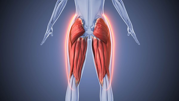
Gluteal compartment syndrome is a rare type of compartment syndrome that is often difficult to identify and/or diagnose. There are three gluteal compartments, and any of the three may be affected by gluteal compartment syndrome. These include the anterior tensor fascia lata compartment, the gluteus medius/minimus compartment, and the gluteus maximus compartment.3
Inadequate perfusion may lead to irreversible damage; thus, early recognition of compartment syndrome is imperative as immediate fasciotomy is required to improve patient outcomes.4 Tissue ischemia from compartment syndrome may lead to rhabdomyolysis. The muscle ischemia and fluid sequestration from compartment syndrome can also lead to hypovolemia, further perpetuating acidemia and acute kidney injury. The sequelae of rhabdomyolysis may include hyperkalemia resulting in life-threatening dysrhythmia, and myoglobinuria resulting in deposition in the distal renal tubules causing tubular cast formation causing acute renal failure.4 Ultimately, this may lead to organ failure or possibly death if not addressed and treated early.
Other potential complications of gluteal compartment syndrome include peripheral neuropathy, complex regional pain syndrome, and sciatic nerve palsy. The sciatic nerve is particularly vulnerable as it may be compressed by compartment syndrome or expanding hematoma leading to potentially irreversible damage or lasting neuropathy/nerve palsy.5 Early diagnosis and fasciotomy are necessary to improve functional prognosis in addition to mitigating morbidity/mortality risks.6
Gluteal compartment syndrome is complicated to diagnose as no definite diagnostic guidelines exist, and it has limited documentation in the literature. As it is largely due to prolonged immobilization, it may be difficult to identify if patients are in a state of altered consciousness or unable to provide history or subjective complaints. One of the preeminent symptoms of compartment syndrome is pain out of proportion to physical examination findings;7 however, patients who cannot provide immediate history are unable to verbalize that they are experiencing unilateral gluteal pain, and thus identifying a tense compartment may be easily missed on initial physical examination. Gluteal compartment syndrome may also be misdiagnosed as a buttock abscess, hematoma, or deep venous thrombosis, which may delay definitive management.8
A thorough secondary examination is imperative in identifying any concerning unilateral extremity findings concerning for compartment syndrome, especially if history is noncontributory as noted above. There are other clues we may use however that may prompt re-evaluation and consideration of compartment syndrome. This includes the development of rhabdomyolysis as indicated by renal dysfunction, acute renal failure, electrolyte derangements, myoglobinuria, rising lactate, decreased sodium bicarbonate levels, leukocytosis, or inflammatory markers. Although many of these serum biomarkers may be within normal ranges, the combination of lab derangements may help prompt thinking about possible compartment syndrome, especially in the setting of a rising CK level as it is a significant indicator of myocyte damage.7
In the following case, we will discuss a 24-year-old male patient who presented to the emergency department after being found unresponsive and in cardiac arrest by EMS, who had return of spontaneous circulation after EMS initiated ACLS protocol. In the emergency department, he remained obtunded requiring intubation and was found to have rhabdomyolysis, metabolic acidosis, and other electrolyte derangements such as hyperkalemia with up-trending serum biomarkers concerning for possible compartment syndrome. Although no significant findings were initially noted on initial secondary examination, these clues ultimately led to the identification of developing gluteal compartment syndrome requiring fasciotomy. Although the patient ultimately recovered from the resulting systemic/metabolic illness, the patient’s functional outcome was impaired by sciatic neuropathy and foot drop/weakness.
CASE REPORT
A 24-year-old male with a past medical history of asthma and substance use disorder presented to the emergency department after return of spontaneous circulation was achieved by EMS. Per EMS, the patient was last seen well by his grandmother in the house the night prior and was found unresponsive the following morning. On EMS arrival at the home, the patient was unresponsive and pulseless. He was noted to have an open needle next to him, vomitus present on his person, and noted to have pinpoint pupils on examination. He was found to be in asystole on paramedic arrival and ACLS was initiated. He was given 16 mg total of intranasal naloxone, and received 3 doses of epinephrine with ongoing CPR. The patient at one point was noted to be in PEA arrest, and subsequent return of spontaneous circulation was achieved. Afterward, the patient was intubated in the field by EMS.
On EMS arrival to the emergency department the patient's heart rate was 131, blood pressure 162/121, intubated and receiving manual respirations, oxygen saturation of 100%. The patient was transferred to a ventilator, respiratory therapy provided suctioning, and the patient was noted to have spontaneous respirations with rhonchorous breath sounds auscultated bilaterally. The patient was noted to appear tremulous, possibly with subtle tonic-clonic movements. He also had pinpoint pupils. As there was concern for possible seizure activity and ongoing overdose, the patient was given an additional 0.4 mg IV naloxone push as well as two 2 mg midazolam pushes, both for possible seizure activity and for additional sedation as the patient was breathing over the ventilator. Given that the patient was hypertensive and there was possible seizure-like activity a propofol drip was started at 20 mcg/kg/min. The patient was initially not febrile on axillary temperature. An i-STAT showed the patient was hyperkalemic, with a potassium of 6.7 and an EKG was immediately obtained which revealed slightly peaked T-waves. The initial pH on i-STAT was 7.0, PCO2 was elevated at 88. POC glucose of 77. Given the hyperkalemia and acidosis, the patient received 1 amp of bicarbonate, 2 g of calcium gluconate, 1 amp of dextrose, and 10 units of insulin. The patient was started on a precedex drip for more adequate sedation. On secondary examination of the patient, no obvious traumatic injuries were noted. CT scan of the head without contrast showed no acute intracranial hemorrhage or mass effect or CT evidence for acute anoxic injury.
Initial labs were significant for leukocytosis with a white count of 29.8 and neutrophilic predominance, hemoglobin 12.5, potassium of 7, anion gap of 25, BUN 26, creatinine 3.1, AST 164, ALT 111, CK 27,000, high-sensitivity troponin 121. The urine toxicology screen was positive for cannabinoids, fentanyl, and cocaine. UA showed 2+ albumin, 4+ glucose, 3+ hemoglobin, 10 WBCs, 8 RBCs, and slight bacteria. One gram of ceftriaxone was given for concern of pneumonia. The patient was then admitted to the medical ICU for further management. Targeted temperature management was initiated with a target temperature of 36C. He was noted to have a tense right lateral gluteal compartment on examination, raising concern for compartment syndrome, particularly as there was evidence of rhabdomyolysis and acute renal failure in his labs. Trauma surgery was consulted due to suspicion of compartment syndrome. The patient was noted to have intermittent generalized tonic-clonic movements, particularly of his bilateral upper extremities, initially thought to be shivering due to targeted temperature management. He was given aspirin and buspirone without effect. The patient continued to have these movements, raising further concern for seizures. He was given 2 mg midazolam, which immediately stopped the movement. His propofol drip was discontinued and he was started on a midazolam drip.
Trauma surgery evaluated the patient and confirmed the suspicion of compartment syndrome. On examination, the right gluteus was firm to touch with overlying skin changes, non-compressible compartments. The patient with myoclonic jerks, on high levels of sedation, could not participate in the exam. The decision was made to bring the patient to the operating room for gluteal fasciotomy.
The patient went to the operating room for a fasciotomy. The deep compartments were tight and bulging and the overlying fascia was opened with satisfactory release of tension. All compartments in the right gluteal area were adequately decompressed. The patient remained intubated and sedated and returned to the MICU. The patient’s hospital course was complicated by rhabdomyolysis with acute kidney injury, compartment syndrome status post fasciotomy right lateral gluteal compartment, and MSSA pneumonia. His course was also complicated by ICU delirium/agitation requiring a precedex drip. On hospital day 5, the patient underwent SBT but failed extubation and required re-intubation due to persistent agitation. He developed MSSA pneumonia, for which he received IV piperacillin/tazobactam. On hospital day 10, he underwent surgical closure of fasciotomy, after which he was able to be extubated and was transitioned to high-flow nasal cannula. The wound VAC was removed on hospital day 13. On hospital day 14, the patient was downgraded to intermediate level of care and ultimately left AMA prior to reevaluation by the trauma surgical service.
The patient presented to the emergency department 2 days later due to right lower extremity weakness/difficulty walking. He was found to be anemic, with a hemoglobin of 5.7 and had a palpable hematoma to fasciotomy site. Trauma surgery was again consulted. The patient underwent a CT scan of the right lower extremity with contrast, which revealed a small elongated hematoma in the soft tissue lateral to the proximal femur at the site of recent surgery.
In the emergency department the patient received 1 unit PRBC transfusion. The patient was booked for operative exploration and hematoma/possible abscess evacuation with trauma surgery, with plans to be admitted to the trauma service afterward. Unfortunately, while waiting to go to the operating room just a few hours after agreeing to surgery and signing his consent for surgery, the patient again left against medical advice.
Two days later, the patient again presented to an outside emergency department with concern for wound dehiscence and worsening lower extremity numbness. He underwent another CT scan showing the hematoma as noted on the previous scan. He also continued to be anemic, with a hemoglobin of 6.6, and received 1 unit PRBC transfusion in the emergency department. Transfer to the main hospital for tertiary-level trauma care was arranged to again be evaluated by trauma surgery. The patient was transferred and admitted to trauma surgery service. The wound was examined at the bedside by the trauma team, and a single suture was removed to allow for evacuation of a large clot just beneath the suture line. His wound was irrigated and the suture was replaced. The wound did not show any secondary signs of infection and the patient’s pain was well controlled. A PM&R consultation was requested for concerns for right foot drop. PM&R recommended outpatient nerve conduction studies and a lightweight AFO boot to help with ambulation. He also received education on wound care so that he could do it himself at home. Given his hemodynamic stability and appropriate pain control, he was deemed safe for discharge 19 days after he first presented to the emergency department.
The patient was seen by PM&R almost 6 months later on an outpatient basis. His neurological exam was consistent with severe left sciatic neuropathy, basically in the peroneal distribution. The prognosis for neurological recovery is unclear at this time but there is no significant recovery thus far at 6 months, so recovery is likely to be far from complete as there is certainly a severe axonal component. EMG/nerve conduction studies were considered for prognostic information but unlikely to change treatment and the patient would likely not be able to tolerate this study at this time. As he had significant neuropathic pain, the patient was given ramp-up instructions for gabapentin. If severe pain persists then the patient will likely be referred to a pain clinic. The patient was given instructions for PT for aggressive range of motion at the right ankle and desensitization. If the patient cannot get good range at the ankle, the patient may be a candidate for Achilles tendon release. The patient was placed on an aggressive home exercise regimen.
DISCUSSION
Early diagnosis of gluteal compartment syndrome can be easily delayed in the altered or unresponsive patient. This diagnosis should be considered by the emergency medicine physician in any patient with prolonged immobilization or another pathogenic mechanism such as trauma or drug overdose. The most frequent symptoms of gluteal compartment syndrome include severe pain, especially with passive motion around the affected buttock, tenseness and/or unilateral swelling.6 However, as noted in this case, without the subjective finding of pain expressed by the patient, this may be easily missed if not considered by the emergency physician. Therefore, the emergency physician needs to consider this diagnosis in any patient with a concerning recent history with other clues such as up-trending serum biomarkers or other evidence of rhabdomyolysis or multiple organ failure if the diagnosis is delayed as noted in this case (demonstrated by Table 1).
Table 1. Metabolic Characteristics
|
Day 1 (ED) 16:00 |
Day 1 (ICU, pre-op) 21:00 |
Day 2 (5 hrs Post-op) |
Day 3 |
Day 7 |
Day 14 (Left AMA) |
|
|
pH |
7.01 |
7.24 |
||||
|
Serum Creatinine |
3.1 |
2.4 |
2.0 |
2.6 |
2.7 |
1.8 |
|
Serum HCO3 |
19 |
16 |
10 |
20 |
23 |
21 |
|
Anion gap |
25 |
22 |
25 |
9 |
12 |
15 |
|
Serum Potassium |
7.0 |
5.0 |
5.0 |
4.1 |
4.3 |
4.7 |
|
Creatine kinase |
27,671 |
59,990 |
62,613 |
33,233 |
2,881 |
|
|
White blood count |
29.8 |
15.1 |
17.9 |
12.6 |
14.4 |
13.7 |
|
AST/ALT |
164/111 |
360/173 |
491/217 |
410/206 |
130,16 |
|
|
Lactate |
10.6 |
9.9 |
13.5 |
1.2 |
1.3 |
It is also imperative to complete a thorough primary and secondary examination of any undifferentiated patient who is altered or unresponsive presenting to the emergency department in order to minimize delay in management of this surgical emergency. As noted with the patient in this case, a delay in the identification of gluteal compartment syndrome may also lead to functional deficits even if the patient recovers from their life-threatening injuries and systemic sequelae.
Gluteal compartment syndrome is largely a clinical diagnosis as there are no standard diagnostic guidelines for this rare condition. If suspected, surgical consultation should not be delayed to try to obtain further imaging or compartment pressures, although this may help in diagnosis or ruling out
other causes of a tense gluteal compartment. CT scans and/or MRIs may help distinguish the extent of muscle edema and/or necrosis as well as any hematoma formation of the affected compartment.6 In addition to early surgical consultation, it is imperative to also manage the systemic sequelae of compartment syndrome, such as fluid resuscitation for rhabdomyolysis and acute renal failure, and to address any concerning electrolyte derangements, although these may also improve after management of the compartment syndrome.
CONCLUSION
Our case demonstrates several rare yet potentially detrimental consequences of drug overdose: respiratory depression resulting in cardiac arrest, prolonged immobilization resulting in gluteal compartment syndrome causing rhabdomyolysis and metabolic derangements, ultimately causing the functional impairment of unilateral sciatic neuropathy and foot drop.9
The emergency physician must have gluteal compartment syndrome on their differential diagnosis in any patient who may have had prolonged immobilization with concerning systemic implications and up-trending serum biomarkers when history is not obtainable directly from the patient to avoid ensuing systemic and functional deficits/permanent disability. When this emergent condition is suspected from clinical evaluation, pathogenic mechanism, and further evaluation with laboratory workup, and possibly imaging/compartment pressures, early surgical consultation for management with fasciotomy should not be delayed.6
REFERENCES
- Panagiotopoulos AC, Vrachnis I, Kraniotis P, Tyllianakis M. Gluteal compartment syndrome following drug-induced immobilization: a case report. BMC Res Notes. 2015;8:35.
- Adib F, Posner AD, O’Hara NN, O’Toole RV. Gluteal compartment syndrome: A systematic review and meta-analysis. Injury. 2021;53(3).
- Jackson JR, Schreyer K. A Pain in the Butt: A Case Series of Gluteal Compartment Syndrome. Clin Pract Cases Emerg Med. 2021;5(2):144-147.
- Tasch JJ, Misodi EO. Delayed Presentation of Acute Gluteal Compartment Syndrome. Am J Case Rep. 2016;17:503-506.
- Rocos B, Ward A. Gluteal compartment syndrome with sciatic nerve palsy caused by traumatic rupture of the inferior gluteal artery: a successful surgical treatment. BMJ Case Rep. 2017;2017:bcr2016216709.
- Iizuka S, Miura N, Fukushima T, Seki T, Sugimoto K, Inokuchi S. Gluteal compartment syndrome due to prolonged immobilization after alcohol intoxication: a case report. Tokai J Exp Clin Med. 2011;36(2):25-28
- Gupta AK, Burgos MI, Lopez-Viego M, Hus N. Gluteal Compartment Syndrome After Prolonged Immobilization in Drug Abusers. Cureus. 2020;12(8):e9847.
- Liu HL, Wong DSY. Gluteal Compartment Syndrome After Prolonged Immobilisation. Asian J Surg. 2009;32(2):123-126.
- Adrish M, Duncalf R, Diaz-Fuentes G, Venkatram S. Opioid overdose with gluteal compartment syndrome and acute peripheral neuropathy. Am J Case Rep. 2014;15:22-26.
- Lederman A, Turk D, Howard A, Reddy S, Stern M. Case study: Gluteal compartment syndrome as a cause of lumbosacral radiculoplexopathy and complex regional pain syndrome. J Rehabil Res Dev. 2016;53(4):483-486.
- Kong GM, Kwon YU, Park JH. Compartment Syndrome of the Gluteus Medius Occurred without Bleeding or Trauma: A Case Report. Hip Pelvis. 2015;27(4):278-282.
- Panagiotopoulos AC, Vrachnis I, Kraniotis P, Tyllianakis M. Gluteal compartment syndrome following drug-induced immobilization: a case report. BMC Res Notes. 2015;8:35.


