Fracture blisters are tense vesicles, or bullae that result from a loss of integrity at the dermal-epidermal junctions secondary to increased interstitial pressure following trauma.1,2
Fluid accumulation combined with the risk of venous stasis following vessel damage contribute to the formation of these blisters. Fracture blisters can present as clear-fluid, or blood-filled lesions, thought to be dependent on the extent of dermal-epidermal separation.3 The most common sites at risk for fracture blister formation are the skin overlying the foot, ankle, tibia shaft, tibial plateau, and elbow.4
Case
A 70-year-old female presented to the emergency department after a 16-hour wilderness evacuation for a trimalleolar fracture and posterior dislocation of the right ankle.
The patient was backpacking when she fell while descending an area of broken, loose rocks. She was unable to ambulate, and without cell phone reception, she waited on the trail until a runner was able to activate search and rescue. She arrived at the ED 18 hours after sustaining her injury.
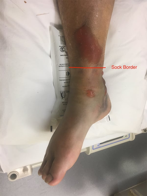
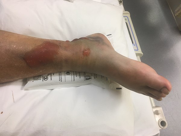
Search and Rescue used a Structural Aluminum Malleable (SAM) splint around the ankle directly on top of the patient’s sock and secured the splint with an ACE wrap. The splint stabilized her injury as she rode a mule, a commonly used method of transport in this region, 8.5 miles along the trail to an ambulance. At the rural ED, the wilderness splint was removed, showing several tense blisters, the largest measuring 5 x 9 cm.
Discussion
The blisters were initially thought to be secondary to inadequate placement of padding under the SAM splint and shear forces of the splint material on the skin. Further inspection, however, showed these to be consistent with fracture blisters in their common anatomic locations (Figure 1).

The SAM splint was a suitable treatment in this situation, but there are several important tips to remember for prehospital injury immobilization. All splints should be well-padded, as bulky dressings can aid in immobilizing the extremity, while controlling swelling via gentle compression.5 Distal pulses should be quickly checked prior to application of any splint materials to achieve a baseline assessment of circulation. Space between the extremity and the splint should be filled with padding to help immobilize the injury and potentially reduce swelling. The splint should provide support proximal and distal to the site of injury. Circulation, motor response, and sensation should be rechecked after application of splint materials and adjusted if there is a change from their initial examination.
Fracture blisters carry an increased risk for infection and are best left intact. They complicate surgical management, and no clear consensus exists on when to surgically repair. When fracture blisters are present preoperatively, there is a higher incidence of postoperative wound infection.2 A detailed skin assessment should be documented and relayed to your consulting orthopedic surgeon.
Outcome
The ankle was reduced in the ED and the patient was transferred the next day to a tertiary center. Orthopedics completed an open reduction internal fixation and required a wound vac to assist with blister management.
References
1. Giordano CP, Scott D, Koval KJ, Kummer F, Atik T, Desai P. Fracture blister formation: a laboratory study. J Trauma. 1995;38(6):907-909.
2. Varela CD, Vaughan TK, Carr JB, Slemmons BK. (1993). Fracture blisters: clinical and pathological aspects. J Orthop Trauma. 1993;7(5):417-427.
3. Strauss EJ, Petrucelli G, Bong M, Koval KJ, Egol KA. Blisters associated with lower-extremity fracture: results of a prospective treatment protocol. J Orthop Trauma. 2006;20(9):618-622.
4. Uebbing CM, Walsh M, Miller JB, Abraham M, Arnold C. Fracture blisters. West J Emerg Med. 2011;12(1):131-133.
5. Tull F, Borrelli Jr J. Soft-tissue injury associated with closed fractures: evaluation and management. J Am Acad Orthop Surg. 2003;11(6):431-438.


