


Case. A middle-aged male with unknown medical history is brought to the emergency department after being found outside on the streets of New York City in February smelling of alcohol and shivering. He is awake but does not respond to questioning. In triage, a temperature could not be obtained.
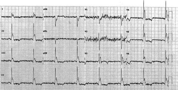
What are you seeing in this ECG?
ANSWER
At first glance, this ECG demonstrates bradycardia which is likely sinus (though artifact makes identification of p-waves difficult); ST-depressions in leads V4-V6; and, most importantly, Osborn waves throughout. An Osborn wave, also known as a J wave, is an ECG finding most often associated with hypothermia. The Osborn wave has the appearance of a camel hump and is a positive deflection between the QRS complex and ST segments of the ECG. The magnitude of the Osborn wave usually correlates directly with the level of hypothermia and so should become less prominent as rewarming occurs.
Other ECG findings that may be seen in hypothermia include bradycardia and prolonged PR, QRS and ST segments. When profound, hypothermia may also precipitate cardiac arrest. A reassuring ECG finding that is present here is the significant artifact seen throughout. This is secondary to shivering, which is desirable in the setting of profound hypothermia. As the patient’s core temperature increases one would see improvement or resolution of the bradycardia, Osborn waves, shivering artifact, and segment prolongation. In a patient who is in cardiac arrest, rewarming may be the trigger for return of circulation. When these ECG findings are present, it is absolutely essential that an accurate temperature be obtained. If hypothermia is identified, decisive steps must be taken to rewarm the patient as this may be a lifesaving intervention.
Learning Points
- The Osborn wave, or J wave, is a positive deflection between the QRS complex and ST segment of an ECG and has a camel hump appearance. It is most often associated with hypothermia.
- The magnitude of the Osborn wave often correlates directly with level of hypothermia and so should improve as rewarming occurs.
- Other ECG findings associated with hypothermia include bradycardia and prolonged PR, QRS and ST segments. Other findings could include asystole or PEA. These ECG changes will improve or resolve with rewarming.