The Patient


A 28-year-old female, who was the restrained front passenger in a motor vehicle accident 1 week before, presents to the ED with abdominal pain. A CT scan of the chest, abdomen and pelvis was negative at the initial visit. On this second visit, she is febrile and has a distended tender abdomen. Her pregnancy test is negative. Point-of-care ultrasound is performed.
What is the diagnosis?
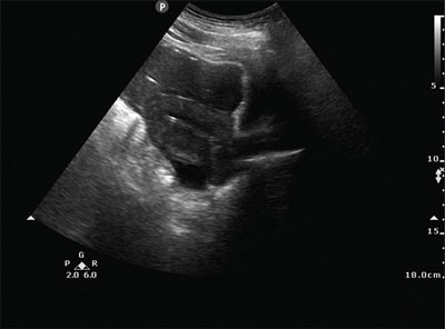
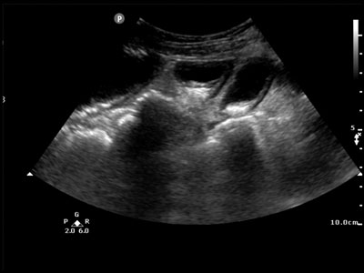
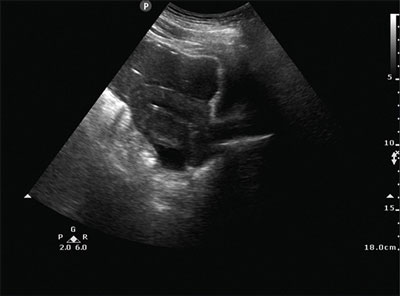
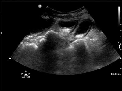
Point-of-care ultrasound of the abdomen demonstrates free fluid in the pelvis (Figure 1) and dilated loops of bowel with absent peristalsis (Figure 2). Confirmatory CT scan revealed a bowel perforation with free fluid and ileus.

Figure 1

Figure 2
Bowel injury secondary to blunt trauma to the abdomen comprises 1% of traumatic injuries, but small bowel is the third most commonly injured abdominal organ after spleen and liver.1-2 Delayed presentation of injuries is even more unusual, with reports in the English language literature limited to case reports and small case series, and is associated with increased mortality.1-10 Patients usually return when initially missed small perforations progress to peritonitis or symptoms of small bowel obstruction.2,6,10 Small tears or hematomas can also heal and lead to strictures and small bowel obstruction days or weeks after a traumatic event.6,9 Though diagnosis is usually made with a CT scan, ultrasound has 80% sensitivity.4Findings on ultrasound include free fluid, dilated loops of bowel with absent peristalsis in the case of obstruction or ileus, focal areas of bowel thickening, and loss of stratification of the small bowel wall.4,11
References
- Kaban G, Somani RA, Carter J. Delayed presentation of small bowel injury after blunt abdominal trauma: case report. J. Trauma. 2004;56(5):1144–1145.
- Fakhry SM, Watts DD, Luchette FA, EAST Multi-Institutional Hollow Viscus Injury Research Group. Current diagnostic approaches lack sensitivity in the diagnosis of perforated blunt small bowel injury: analysis from 275,557 trauma admissions from the EAST multi-institutional HVI trial. J. Trauma.2003;54(2):295–306.
- Bouliaris K, Karangelis D, Spanos K, Germanos S, Alexiou E, Giaglaras A. Ileosigmoid fistula and delayed ileal obstruction secondary to blunt abdominal trauma: A case report. J. Med. Case Reports.2011;5:507.
- Shih HC, Liu M, Wu JK, et al. Blunt gastrointestinal injury in trauma patients. Am J Emerg Med.1995;13(1):82–84.
- McGuigan A, Brown R. Early and delayed presentation of traumatic small bowel injury. BMJ Case Rep.2016.
- Ekeh AP, Saxe J, Walusimbi M, et al. Diagnosis of blunt intestinal and mesenteric injury in the era of multidetector CT technology – are results better? J. Trauma. 2008;65(2):354–359.
- Pikoulis E, Delis S, Psalidas N, et al. Presentation of blunt small intestinal and mesenteric injuries. Ann R Coll Surg Engl. 2000;82(2):103–106.
- Nosanov LB, Barthel ER, Pierce JR. Sigmoid perforation and bucket-handle tear of the mesocolon after bicycle handlebar injury: a case report and review of the literature. J Pediatr Surg.2011;46(12):e33-35.
- Hamidian JA, Johnson L, Youssef AM. Delayed small bowel perforation following blunt abdominal trauma: A case report and review of the literature. Asian J Surg. 2016;39(2):109–112.
- Subramanian V, Raju RS, Vyas FL, Joseph P, Sitaram V. Delayed jejunal perforation following blunt abdominal trauma. Ann R Coll Surg Engl. 2010;92(2):W23-24.
- Chang YS, Wang HP, Huang GT, Chen SC, Wang SM. Sonographic detection of delayed small bowel perforations after blunt abdominal trauma. J Clin Ultrasound. 2000;28(3):142–145.


