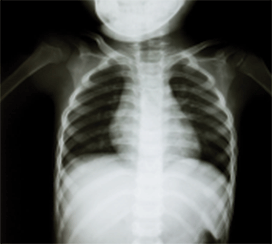
From the November 2013 issue of Pediatric Emergency Medicine Practice, “Emergency Management Of Blunt Chest Trauma In Children: An Evidence-Based Approach.” Reprinted with permission. To access your EMRA member benefit of free online access to all EM Practice, Pediatric EM Practice, and EM Practice Guidelines Update issues, go to www.ebmedicine.net/emra, call 1-800-249-5770, or email ebm@ebmedicine.net.
- “The infant presented with shortness of breath. There was no fever, the oxygen saturation was normal, and the lungs were clear. I thought it was respiratory syncytial virus. I didn't think it could be from abusive chest trauma.”
Infants with abusive head trauma and/or abusive chest trauma can present with nonspecific signs and symptoms, including respiratory complaints. These patients may present similarly to infants with bronchiolitis. Emergency clinicians should look for the presence of upper respiratory symptoms, runny nose, fevers, etc, that may indicate an upper respiratory infection or bronchiolitis. Absence of these symptoms should generate a high index of suspicion to evaluate for potential nonaccidental trauma. - “The 7-year-old presented with confusion and a femur fracture. His CT scan showed a splenic laceration. I didn't think his persistent hypotension was caused by a cardiac tamponade.”
Blunt cardiac injury is usually seen in a patient with multisystem injuries. Emergency clinicians may be distracted when seeing additional injuries (such as intra-abdominal pathology or extremity fractures). It is important to consider blunt cardiac injury when assessing the child with multisystem injury. - “The 4-year-old boy came in after a high-speed car accident. He was not belted and was thrown into the windshield. His chest x-ray revealed multiple rib fractures and a pneumothorax. He was admitted to the pediatric intensive care unit, where they subsequently found a traumatic aortic dissection. I can't believe I forgot to look at his mediastinum on the chest x-ray! It was definitely widened. I didn't think children could have this injury.”
Emergency clinicians should carefully examine the chest x-ray for abnormalities such as widened mediastinum or abnormal apical knob. Although aortic injuries are uncommon in the pediatric patient, they do occur. - “The 8-year-old was riding her bike when she was struck by a car. Her initial heart rate was 140 beats/min with a blood pressure of 70/30 mm Hg. She presented with a GCS of 10 and was immediately intubated. Chest x-ray revealed rib fractures. Her FAST exam was negative, and CT of chest and abdomen was negative for acute injury. She remained tachycardic and intermittently hypotensive. What injury am I missing with a negative pan scan?”
This young girl was later diagnosed with a cardiac contusion, the most common form of blunt cardiac injury. Clues to her diagnosis were tachycardia, rhythm abnormalities, and hypotension. An ECG and troponin may have helped with the diagnosis. - “The 5-year-old girl presented after a large TV fell on her chest. She arrived tachycardic with a blood pressure of 100/65 mm Hg. Her initial oxygen saturation was 99% and her chest x-ray revealed only 2 rib fractures. Three hours later, she developed respiratory distress and became hypoxic, with an oxygen saturation of 82%. I had to intubate her. Repeat chest x-rays revealed a large pulmonary contusion. How did I miss it?”
Findings of pulmonary contusion may be delayed for several hours in the pediatric patient. Respiratory distress and hypoxia may develop after an initial chest x-ray that is normal. The emergency clinician should be aware of delayed clinical findings from a developing pulmonary contusion. Initial findings to suggest a developing pulmonary contusion includes relative hypoxia, with saturations in the 93% to 95% range. - “We came to the scene of a 7-year-old with head, chest, and abdominal trauma. She was breathing OK with a saturation of 97%. Looking back at the record, I can't believe we spent 44 minutes on the scene. We should have transported faster.”
Prehospital delay of transport should be avoided as much as possible, especially if the delay is from repeated intravenous or intubation attempts. Emphasis should be placed on oxygenation, ventilation, treatment of tension pneumothorax, and motion restriction. Specific attention to oxygenation/ventilation is the first priority, and this can usually be accomplished with manual airway maneuvers; intubation is rarely required for pediatric patients. - “The 2-year-old boy presented with a high respiratory rate, hypotension, head injury with altered mental status, and a femur fracture. I was distracted by his other injuries. I can't believe I missed the first-rib fracture, cardiac contusion, and small aortic tear.”
A high index of suspicion is needed to diagnose chest trauma. External injuries may not be immediately evident, and other organ systems may distract the emergency clinician. Holmes, et al., found 6 clinical findings that helped to predict chest injuries. These include: (1) abnormal chest auscultation, (2) low systolic blood pressure, (3) GCS <15, (4) abnormal thoracic examination, (5) elevated respiratory rate, and (6) femur fracture.


