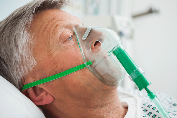
A 56-year-old male with a history of end stage COPD, sarcoidosis, and pulmonary hypertension comes into your ED in respiratory failure. His initial oxygen saturation is 38% on 6L nasal cannula. You quickly place him on BiPAP and his oxygen saturation jumps up to 74%. However, he becomes increasingly drowsy and more difficult to arouse, despite nebulized medications and IV steroids. His first ABG reveals a pH of 7.23, a pCO2 >100, and a pO2 of 58.
Standard medical treatment
We have come a long way in the treatment of acute hypercapnic respiratory failure. No longer do we intubate every wheezing hypoxic and tachypneic patient who comes through the door. Like with all patients, the standard of care is to begin with the ABCs. Initial management should include oxygen supplementation either via high flow humidified oxygen (HFHO) or nasal cannula/non-rebreather mask, simultaneously with nebulized albuterol, IV steroid medication ± magnesium, and antibiotics (if necessary). HFHO therapy has been shown to improve oxygenation and provide a small amount of PEEP in these patients, and may also decrease the need for intubation. If HFHO is not available, it is important to leave a nasal cannula on the patient at all times, even underneath the non-rebreather mask. This serves to prolong adequate oxygenation during the apneic period of intubation after the non-rebreather mask is taken off.1,2
Non-invasive positive pressure ventilation (NIPPV)
Numerous research articles have substantiated NIPPV to be the most effective first-line therapy when clinical signs (tachypnea, accessory muscle use, acidosis) of hypercapneic respiratory failure persist despite standard medical treatment. In patients with the right history and physical exam findings, BiPAP is often initiated immediately, in conjunction with other standard therapies. NIPPV has been found to reduce mortality, help avoid endotracheal intubation, and decrease treatment failure when initiated early, and before the onset of severe acidosis.3
The pH is the most significant value for predicting NIPPV failure.4 One study revealed an association between worsening pH within the first hour of treatment and NIPPV failure.5 This suggests a “golden hour” in which BiPAP should improve the level of acidosis, or the patient is very likely to develop progressive respiratory failure and require intubation. However, NIPPV should not be applied indiscriminately – there are clear-cut limitations of efficacy in patients with isolated hypoxic respiratory failure and/or severe acidosis. It remains controversial for treatment of status asthmaticus, ARDS, severe pulmonary hypertension, or when there is clear evidence of clinical decompensation. Using NIPPV in these patient populations may delay necessary intubation and result in a potentially avoidable death.6,7
Severe acidosis
Just how acidotic must a patient be to require intubation? One study found no significant difference in mortality between a group with a pH of 7.2-7.25 and a group with a pH of 7.26-7.3 when treated with NIPPV.8 There is a school of thought that suggests that in these patients, a pH
< 7.25 necessitates immediate intubation. However, clinical judgment will always be the superior guideline to follow, regardless of the numbers.
Post-intubation ventilation settings should be specifically tailored to each individual patient and should be adjusted according to ABG results. A repeat blood gas should be obtained within 10-15 minutes of being placed on the ventilator. Tenuous patients are likely to require an arterial catheter for future ABG draws.9 If there is difficulty in obtaining an ABG, the venous pH can be multiplied by a factor of 1.004 to accurately predict arterial pH.10
Case closure
Since he was not improving on BiPAP, you intubate the patient. His oxygen saturation immediately rises to 100%, and his post-intubation ABG reveals a normal pH, and a pCO2 of 55. He is admitted to the ICU and is eventually extubated and discharged home several days later.
References
- Sztrymf B, et al. Beneficial effects of humidified high flow nasal oxygen in critical care patients: a prospective pilot study. Intensive Care Medicine. 2011 Nov;17(11):1780-1786
- Walls RM, Murphy MF. “Manual of Emergency Airway Management” Pharmacology and Techniques. Fourth Edition. Lippincott Williams & Wilkins, 2012. 222-224. Print.
- Lightowler JV, et al. Non-invasive positive pressure ventilation to treat respiratory failure resulting from exacerbations of chronic obstructive pulmonary disease: Cochrane systematic review and meta-analysis. British Medical Journal. 2003 Jan;326(185).
- Thomas A, et al. The Strength of Association Between the Risk of Endotracheal Intubtation and Initial Arterial Blood (ABG) pH in Patients Presenting with Acute Respiratory Failure (AHRF) Treated with Non-invasive Ventilation. Thorax. 2010 Dec;65(4):P158.
- Baglioni S, et al. Predictors of Noninvasive Mechanical Ventilation Failure or Success in Patients With Severe Respiratory Acidosis. CHEST. 2011 Oct;140:399A.
- Aboussouan LS, Ricaurte B. Noninvasive positive pressure ventilation: Increasing use in acute care. Cleveland Clinic Journal of Medicine. 2010 May;77(5):307-316.
- Parola D, et al. Treatment of acute exacerbations with non-invasive ventilation in chronic hypercapnic COPD patients with pulmonary hypertension. European Review for Medical and Pharmacological Sciences. 2012 Feb;16(2):183-191.
- Thomas A, et al. Non-invasive Ventilation (NIV) in Chronic Obstructive Pulmonary Disease (COPD) Exacerbations with Acute Hypercapnic Respiratory Failure (AHRF) with pH. Thorax. 2010 Dec;65(4):S70.
- Kovacs G, Green RS. “Postintubation Management” Airway Management in Emergencies. Second Edition. Shelton: People's Medical Publishing House-USA, 2011. 230. Print.
- Ak A, et al. Prediction of Arterial Blood Gas Values from Venous Blood Gas Values in Patients with Acute Exacerbations of Chronic Obstructive Pulmonary Disease. The Tohoku Journal of Experimental Medicine. 2006 Dec;210(4):285-290.


