


CASES
Case 1. The Patient
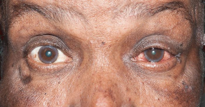
A 61-year-old female complains of sudden onset right eye pain, headache, blurred vision, and vomiting 2 hours prior to coming to the ED. Physical examination reveals a patient who is alert and in pain. Vital signs are normal. Visual acuity: Can count fingers at three feet OD, 20/30 OS. Extraocular movements are normal. See image below for additional eye exam findings.

What is the diagnosis?
Case 2. The Patient
A 65-year-old male with sarcoidosis complains of gradual onset left eye pain and photophobia. These symptoms have occurred previously. Consensual and direct photophobia is present. Physical examination reveals a patient who is alert and in pain. Vital signs are normal. Visual acuity: 20/25 OD, 20/200 OS. Intraocular pressure is 13 mmHg OD, 16 mmHg OS by tonopen. Extraocular movements are normal. White blood cells are seen in the anterior chamber of the left eye on slit lamp examination. See image below for additional eye exam findings.
What is the diagnosis?
Case 1. The Diagnosis.
Acute angle-closure glaucoma.
The patient's nonreactive midrange dilated right pupil, irregular cornea, conjunctival injection, and elevated IOP with eye pain, headache, blurred vision and vomiting strongly suggests acute angle-closure glaucoma (ACG). Intraocular pressures (IOP) were measured at 52 mmHg OD and 19 mmHg OS. ED treatment of ACG includes immediate ophthalmology consultation and administering medications to decrease aqueous humor production and increase aqueous outflow, and administering osmotic agents to decrease intraocular pressure.
Case 2. The Diagnosis.
Acute iritis or uveitis.
Inflammation of the anterior uvea (iris and ciliary body) is synonymous with acute iritis. Iritis is manifest by eye redness and pain, photophobia, miosis, limbic injection, and inflammatory cells and/or protein accumulation (“flare”) in the anterior chamber. Treatment depends on the cause. Common causes include traumatic iritis and infectious causes such as bacterial keratitis, CMV, tuberculosis, toxoplasmosis, syphilis, and cat-scratch disease. Inflammatory diseases with associated iritis include the spondyloarthitides, sarcoidosis, Behçet's disease, SLE, Crohn's disease, and ulcerative colitis. Ophthalmology consultation is needed to manage the ocular complications of iritis while the etiology is determined. Iritis in this patient was secondary to his sarcoidosis.